
Search Count: 17
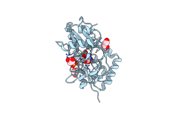 |
Structure Of Glua2-N775S Ligand-Binding Domain (S1S2J) In Complex With Glutamate And Rubidium Bromide At 1.75 A Resolution
Organism: Rattus norvegicus
Method: X-RAY DIFFRACTION Resolution:1.75 Å Release Date: 2019-05-15 Classification: MEMBRANE PROTEIN Ligands: GLU, SO4, GOL, BR |
 |
Structure Of Glua2O Ligand-Binding Domain (S1S2J) In Complex With Glutamate And Sodium Bromide At 1.95 A Resolution
Organism: Rattus norvegicus
Method: X-RAY DIFFRACTION Resolution:1.95 Å Release Date: 2019-05-15 Classification: MEMBRANE PROTEIN Ligands: GLU, BR, GOL, NA, ACT |
 |
Crystal Structure Of The Glua2 Ligand-Binding Domain (S1S2J) In Complex With (S)-2-Amino-3-(5-(2-(3-(Aminomethyl)Benzyl)-2H-Tetrazol-5-Yl)-3-Hydroxyisoxazol-4-Yl)Propanoic Acid At Resolution 1.55 A Resolution
Organism: Rattus norvegicus
Method: X-RAY DIFFRACTION Resolution:1.55 Å Release Date: 2016-03-02 Classification: MEMBRANE PROTEIN Ligands: SO4, 5XP, GOL, ACT, EDO, LI |
 |
Crystal Structure Of The Glua2 Ligand-Binding Domain (S1S2J) In Complex With (S)-2-Amino-3-(5-(2-(3-Methylbenzyl)-2H-Tetrazol-5-Yl)-3-Hydroxyisoxazol-4-Yl)Propanoic Acid At 1.6 A Resolution
Organism: Rattus norvegicus
Method: X-RAY DIFFRACTION Resolution:1.60 Å Release Date: 2016-03-02 Classification: MEMBRANE PROTEIN Ligands: SO4, GOL, ACT, 5XO, EDO |
 |
Crystal Structure Of The Glua2 Ligand-Binding Domain (S1S2J) In Complex With (S)-2-Amino-3-(5-(2-(3-Chlorobenzyl)-2H-Tetrazol-5-Yl)-3-Hydroxyisoxazol-4-Yl)Propanoic Acid At 2.3 A Resolution
Organism: Rattus norvegicus
Method: X-RAY DIFFRACTION Resolution:2.30 Å Release Date: 2016-03-02 Classification: MEMBRANE PROTEIN Ligands: 5XN, SO4, GOL, CL, EDO, PG4 |
 |
Organism: Leishmania major
Method: X-RAY DIFFRACTION Resolution:2.40 Å Release Date: 2015-05-27 Classification: TRANSFERASE Ligands: T5A, ZN |
 |
Organism: Leishmania major
Method: X-RAY DIFFRACTION Resolution:2.74 Å Release Date: 2015-05-27 Classification: TRANSFERASE Ligands: PO4, THM, ZN |
 |
Organism: Leishmania major
Method: X-RAY DIFFRACTION Resolution:3.00 Å Release Date: 2015-05-27 Classification: TRANSFERASE Ligands: ZN, TTP, MG |
 |
Organism: Plasmodium falciparum
Method: X-RAY DIFFRACTION Resolution:2.40 Å Release Date: 2012-08-29 Classification: HYDROLASE/HYDROLASE INHIBITOR Ligands: DUA, GOL |
 |
Organism: Plasmodium falciparum 3d7, Escherichia coli
Method: X-RAY DIFFRACTION Resolution:1.65 Å Release Date: 2012-08-29 Classification: HYDROLASE/HYDROLASE INHIBITOR Ligands: DU3, SO4 |
 |
Organism: Plasmodium falciparum, Synthetic construct
Method: X-RAY DIFFRACTION Resolution:2.60 Å Release Date: 2012-08-29 Classification: HYDROLASE/HYDROLASE INHIBITOR Ligands: DU2, SO4 |
 |
Organism: Plasmodium falciparum, Synthetic construct
Method: X-RAY DIFFRACTION Resolution:1.80 Å Release Date: 2012-08-29 Classification: HYDROLASE/HYDROLASE INHIBITOR Ligands: DU4, SO4 |
 |
The Crystal Structure Of Leishmania Major Dutpase In Complex With Substrate Analogue Dupnpp
Organism: Leishmania major
Method: X-RAY DIFFRACTION Resolution:1.86 Å Release Date: 2011-03-16 Classification: HYDROLASE Ligands: DUP, CA |
 |
Organism: Leishmania major
Method: X-RAY DIFFRACTION Resolution:2.40 Å Release Date: 2011-03-16 Classification: HYDROLASE Ligands: UMP, SO4, MG |
 |
Organism: Leishmania major
Method: X-RAY DIFFRACTION Resolution:2.28 Å Release Date: 2011-03-16 Classification: HYDROLASE Ligands: DUR, SO4 |
 |
The Crystal Structure Of A Complex Of Leishmania Major Dutpase With Substrate Analogue Dupnhp
Organism: Leishmania major
Method: X-RAY DIFFRACTION Resolution:2.34 Å Release Date: 2007-04-17 Classification: HYDROLASE Ligands: DUN, MG |
 |
The Crystal Structure Of A Complex Of Campylobacter Jejuni Dutpase With Substrate Analogue Dupnhpp
Organism: Campylobacter jejuni
Method: X-RAY DIFFRACTION Release Date: 2007-03-27 Classification: HYDROLASE Ligands: DUP, MG |