
Search Count: 20
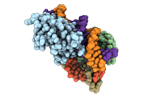 |
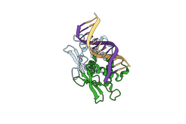
Co-Crystal Structure Of Yeast Forkhead Transcription Factor Fkh1 Bound To Dna
Organism: Saccharomyces cerevisiae, Synthetic construct
Method: X-RAY DIFFRACTION Release Date: 2025-09-24 Classification: DNA BINDING PROTEIN/DNA Ligands: K |
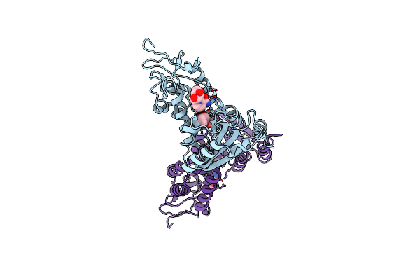 |
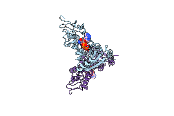
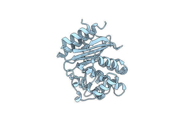
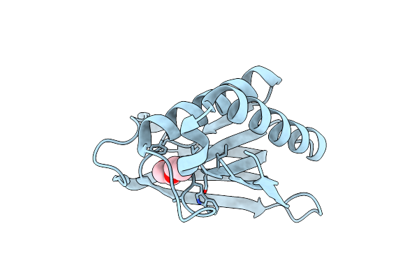
Organism: Homo sapiens
Method: X-RAY DIFFRACTION Resolution:2.10 Å Release Date: 2024-11-06 Classification: HYDROLASE/INHIBITOR Ligands: NNR, RIB |
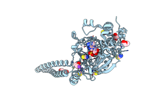 |
An Orthogonal Seryl-Trna Synthetase/Trna Pair For Noncanonical Amino Acid Mutagenesis In Escherichia Coli
Organism: Methanosarcina mazei (strain atcc baa-159 / dsm 3647 / goe1 / go1 / jcm 11833 / ocm 88)
Method: X-RAY DIFFRACTION Resolution:1.45 Å Release Date: 2020-11-04 Classification: LIGASE Ligands: SSA, PEG, DMS, SCN, K, CL |
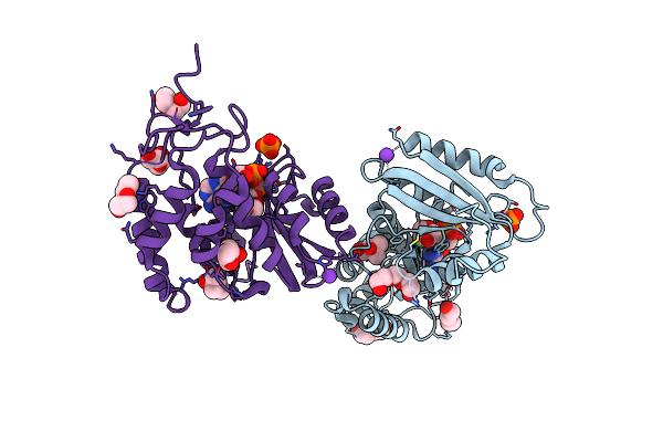 |
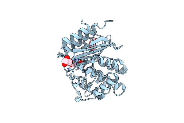
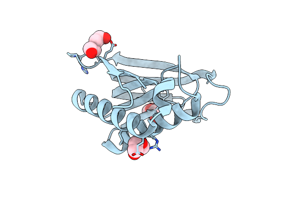
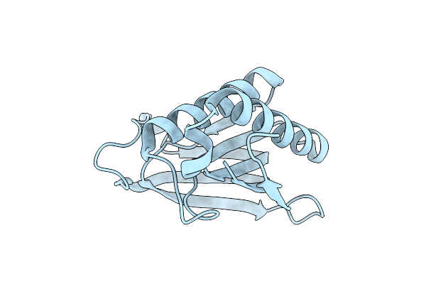
Organism: Homo sapiens
Method: X-RAY DIFFRACTION Resolution:1.50 Å Release Date: 2020-06-03 Classification: HYDROLASE Ligands: ROJ, K, PO4, PEG, MPD, PGE |
 |
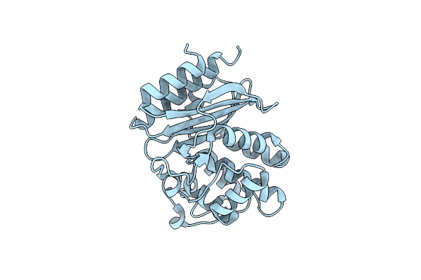
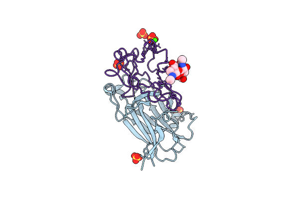
Organism: Homo sapiens
Method: X-RAY DIFFRACTION Resolution:2.40 Å Release Date: 2018-11-21 Classification: HYDROLASE/HYDROLASE INHIBITOR Ligands: ZNA |
 |
E. Coli Chorismate Mutase With Orthogonal Interface Containing P-Benzoyl Phenylalanine
Organism: Escherichia coli
Method: X-RAY DIFFRACTION Resolution:2.00 Å Release Date: 2017-05-10 Classification: ISOMERASE |
 |
Crystal Structure Of P-Acrylamido-Phenylalanine Modified Tem1 Beta-Lactamase From Escherichia Coli : V216Acrf Mutant
Organism: Escherichia coli
Method: X-RAY DIFFRACTION Resolution:1.54 Å Release Date: 2015-05-20 Classification: HYDROLASE Ligands: GOL, MES, PEG |
 |
Crystal Structure Of P-Acrylamido-Phenylalanine Modified Tem1 Beta-Lactamase From Escherichia Coli :E166N Mutant
Organism: Escherichia coli
Method: X-RAY DIFFRACTION Resolution:1.80 Å Release Date: 2015-05-20 Classification: HYDROLASE |
 |
Crystal Structure Of Cephalexin Bound Acyl-Enzyme Intermediate Of Val216Acrf Mutant Tem1 Beta-Lactamase From Escherichia Coli: E166N And V216Acrf Mutant.
Organism: Escherichia coli
Method: X-RAY DIFFRACTION Resolution:1.70 Å Release Date: 2015-05-20 Classification: HYDROLASE |
 |
Biphenylalanine Modified Threonyl-Trna Synthetase From Pyrococcus Abyssi: I11Bif, F42W, Y79A, And F123Y Mutant
Organism: Pyrococcus abyssi ge5
Method: X-RAY DIFFRACTION Resolution:1.95 Å Release Date: 2015-03-18 Classification: LIGASE Ligands: PEG |
 |
Biphenylalanine Modified Threonyl-Trna Synthetase From Pyrococcus Abyssi: I11Bif, Y79I, And F123A Mutant
Organism: Pyrococcus abyssi ge5
Method: X-RAY DIFFRACTION Resolution:2.05 Å Release Date: 2015-03-18 Classification: LIGASE |
 |
Biphenylalanine Modified Threonyl-Trna Synthetase From Pyrococcus Abyssi: 11Bif, 42F, 79S, And 123A Mutant
Organism: Pyrococcus abyssi ge5
Method: X-RAY DIFFRACTION Resolution:2.36 Å Release Date: 2015-03-18 Classification: LIGASE |
 |
Biphenylalanine Modified Threonyl-Trna Synthetase From Pyrococcus Abyssi: 11Bif, 42F, 79S, And 123V Mutant
Organism: Pyrococcus abyssi ge5
Method: X-RAY DIFFRACTION Resolution:2.10 Å Release Date: 2015-03-18 Classification: LIGASE |
 |
Biphenylalanine Modified Threonyl-Trna Synthetase From Pyrococcus Abyssi: 11Bif, 42F, 79V, And 123A Mutant
Organism: Pyrococcus abyssi ge5
Method: X-RAY DIFFRACTION Resolution:2.10 Å Release Date: 2015-03-18 Classification: LIGASE Ligands: PEG |
 |
Biphenylalanine Modified Threonyl-Trna Synthetase From Pyrococcus Abyssi: I11Bif, Y79V, And F123V Mutant
Organism: Pyrococcus abyssi ge5
Method: X-RAY DIFFRACTION Resolution:2.50 Å Release Date: 2015-03-18 Classification: LIGASE |
 |
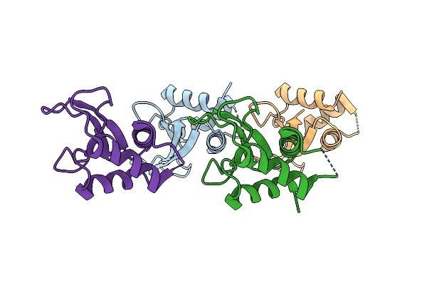
Structure Of The Human Epha3 Receptor Ligand Binding Domain Complexed With Ephrin-A5
Organism: Homo sapiens
Method: X-RAY DIFFRACTION Resolution:2.26 Å Release Date: 2014-06-11 Classification: TRANSFERASE/TRANSFERASE RECEPTOR Ligands: SO4, CA |
 |
The Crystal Structure Of The Dna Binding Domain Of Virf-1 From The Oncogenic Kshv
Organism: Human herpesvirus 8
Method: X-RAY DIFFRACTION Resolution:2.38 Å Release Date: 2013-03-13 Classification: DNA BINDING PROTEIN |
 |
The Complex Crystal Structure Of The Dna Binding Domain Of Virf-1 From The Oncogenic Kshv With Dna
Organism: Human herpesvirus 8, Synthetic construct
Method: X-RAY DIFFRACTION Resolution:1.48 Å Release Date: 2013-03-13 Classification: DNA BINDING PROTEIN/DNA |
 |
Organism: Homo sapiens
Method: X-RAY DIFFRACTION Resolution:2.70 Å Release Date: 2006-02-07 Classification: SIGNALING PROTEIN |
 |
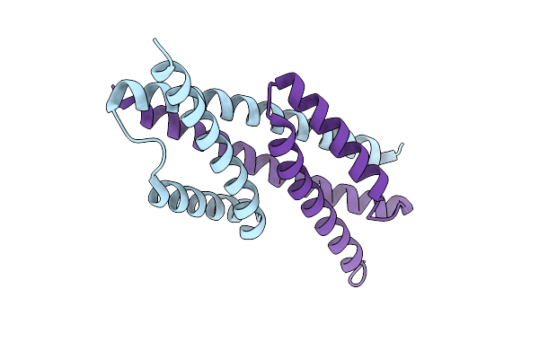
Organism: Rattus norvegicus
Method: SOLUTION NMR Release Date: 2005-04-05 Classification: CELL ADHESION |