
Search Count: 129
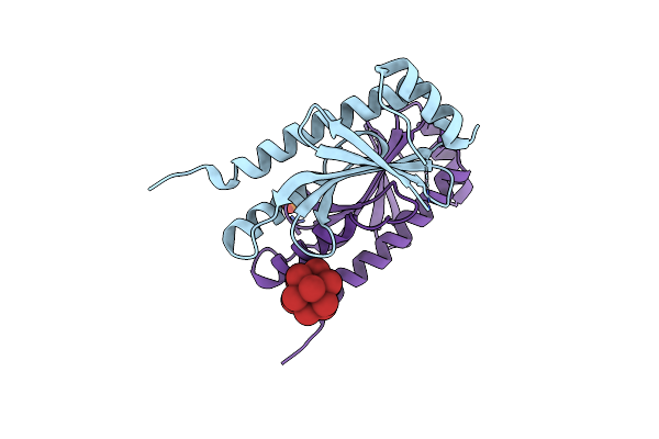 |
Crystal Structure Of The Type Iii Secretion Chaperone Veca From Vibrio Parahaemolyticus
Organism: Vibrio parahaemolyticus
Method: X-RAY DIFFRACTION Release Date: 2025-06-25 Classification: CHAPERONE Ligands: PO4, TBR |
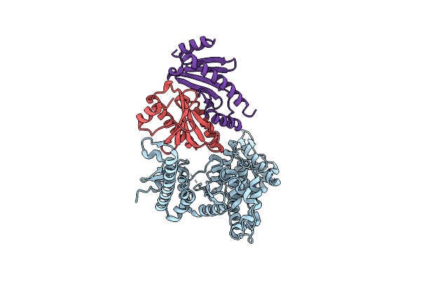 |
Crystal Structure Of The Virulence Effector Vepa In Complex With Its Secretion Chaperone Veca
Organism: Vibrio parahaemolyticus
Method: X-RAY DIFFRACTION Release Date: 2025-06-25 Classification: TOXIN |
 |
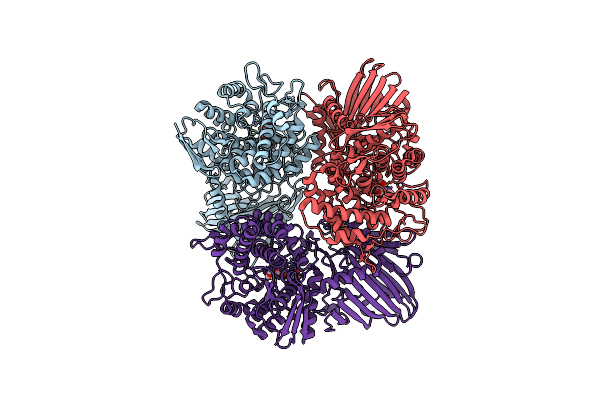
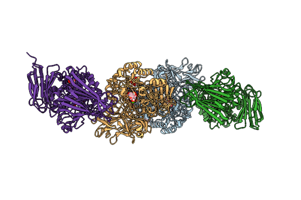
Cryo-Em Structure Of The Type Ivb Pilus From Enterotoxigenic Escherichia Coli
Organism: Escherichia coli
Method: ELECTRON MICROSCOPY Release Date: 2025-05-28 Classification: CELL ADHESION |
 |
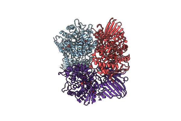
Cryo-Em Structure Of The Type I Pilus From Enterotoxigenic Escherichia Coli
Organism: Escherichia coli
Method: ELECTRON MICROSCOPY Release Date: 2025-05-28 Classification: CELL ADHESION |
 |
Crystal Structure Of Gh65 Alpha-1,2-Glucosidase From Flavobacterium Johnsoniae In Complex With 1-Deoxynojirimycin
Organism: Flavobacterium johnsoniae uw101
Method: X-RAY DIFFRACTION Resolution:1.90 Å Release Date: 2025-03-19 Classification: HYDROLASE Ligands: NOJ, EDO |
 |
Crystal Structure Of Gh65 Alpha-1,2-Glucosidase From Flavobacterium Johnsoniae In Complex With Castanospermine
Organism: Flavobacterium johnsoniae uw101
Method: X-RAY DIFFRACTION Resolution:1.60 Å Release Date: 2025-03-19 Classification: HYDROLASE Ligands: CTS, EDO |
 |
Crystal Structure Of The Receptor Binding Domain Of Sars-Cov-2 Spike Protein In Complex With Ce9
Organism: Severe acute respiratory syndrome coronavirus 2, Synthetic construct
Method: X-RAY DIFFRACTION Release Date: 2025-01-15 Classification: VIRAL PROTEIN/INHIBITOR Ligands: NAG, GOL |
 |
Crystal Structure Of The Receptor Binding Domain Of Sars-Cov-2 Omicron Ba.2 Variant Spike Protein In Complex With Ce149
Organism: Severe acute respiratory syndrome coronavirus 2, Synthetic construct
Method: X-RAY DIFFRACTION Release Date: 2025-01-15 Classification: VIRAL PROTEIN/INHIBITOR Ligands: GOL |
 |
Crystal Structure Of The Receptor Binding Domain Of Sars-Cov-2 Alpha Variant Spike Protein In Complex With Ce59
Organism: Severe acute respiratory syndrome coronavirus 2, Synthetic construct
Method: X-RAY DIFFRACTION Release Date: 2025-01-15 Classification: VIRAL PROTEIN/INHIBITOR Ligands: NAG |
 |
Crystal Structure Of The Receptor Binding Domain Of Sars-Cov-2 Delta Variant Spike Protein In Complex With Ce59
Organism: Severe acute respiratory syndrome coronavirus 2, Synthetic construct
Method: X-RAY DIFFRACTION Release Date: 2025-01-15 Classification: VIRAL PROTEIN/INHIBITOR Ligands: GOL |
 |
Crystal Structure Of The Receptor Binding Domain Of Sars-Cov-2 Alpha Variant Spike Protein In Complex With Ce41
Organism: Severe acute respiratory syndrome coronavirus 2, Synthetic construct
Method: X-RAY DIFFRACTION Release Date: 2025-01-15 Classification: VIRAL PROTEIN/INHIBITOR Ligands: NAG |
 |
Crystal Structure Of The Receptor Binding Domain Of Sars-Cov-2 Omicron Ba.2 Variant Spike Protein In Complex With Cespiace
Organism: Severe acute respiratory syndrome coronavirus 2, Synthetic construct
Method: X-RAY DIFFRACTION Release Date: 2025-01-15 Classification: VIRAL PROTEIN/INHIBITOR Ligands: GOL |
 |
Crystal Structure Of The Receptor Binding Domain Of Sars-Cov-2 Omicron Ba.5 Variant Spike Protein In Complex With Cespiace
Organism: Severe acute respiratory syndrome coronavirus 2, Synthetic construct
Method: X-RAY DIFFRACTION Release Date: 2025-01-15 Classification: VIRAL PROTEIN/INHIBITOR Ligands: GOL |
 |
Crystal Structure Of The Receptor Binding Domain Of Sars-Cov-2 Omicron Xbb.1.5 Variant Spike Protein In Complex With Cespiace
Organism: Severe acute respiratory syndrome coronavirus 2, Synthetic construct
Method: X-RAY DIFFRACTION Release Date: 2025-01-15 Classification: VIRAL PROTEIN/INHIBITOR Ligands: GOL, NA |
 |
Cryo-Em Structure Of Sars-Cov-2 Spike Ectodomain (Hexapro, Omicron Ba.2 Variant) In Complex With Cespiace
Organism: Severe acute respiratory syndrome coronavirus 2, Synthetic construct
Method: ELECTRON MICROSCOPY Release Date: 2025-01-15 Classification: VIRAL PROTEIN/INHIBITOR Ligands: NAG |
 |
Cryo-Em Structure Of Sars-Cov-2 Spike Ectodomain (Hexapro, Omicron Ba.5 Variant) In Complex With Cespiace, Class 1
Organism: Severe acute respiratory syndrome coronavirus 2, Synthetic construct
Method: ELECTRON MICROSCOPY Release Date: 2025-01-15 Classification: VIRAL PROTEIN/INHIBITOR Ligands: NAG |
 |
Cryo-Em Structure Of Sars-Cov-2 Spike Ectodomain (Hexapro, Omicron Ba.5 Variant) In Complex With Cespiace, Class 2
Organism: Severe acute respiratory syndrome coronavirus 2, Synthetic construct
Method: ELECTRON MICROSCOPY Release Date: 2025-01-15 Classification: VIRAL PROTEIN/INHIBITOR Ligands: NAG |
 |
Activity-Stability Trade-Off Observed In Variants At Position 315 Of The Gh10 Xylanase Xynr
Organism: Bacillus sp. tar1
Method: X-RAY DIFFRACTION Resolution:1.90 Å Release Date: 2024-05-22 Classification: HYDROLASE Ligands: CA, PEG |
 |
Organism: Bacillus sp. tar1
Method: X-RAY DIFFRACTION Resolution:1.80 Å Release Date: 2024-05-22 Classification: HYDROLASE Ligands: CA, ACT, MPD, TRS, PEG |
 |
Crystal Structure Of Gh97 Glucodextranase From Flavobacterium Johnsoniae In Complex With Glucose
Organism: Flavobacterium johnsoniae (strain atcc 17061 / dsm 2064 / jcm 8514 / nbrc 14942 / ncimb 11054 / uw101)
Method: X-RAY DIFFRACTION Resolution:1.95 Å Release Date: 2024-05-08 Classification: HYDROLASE Ligands: CA, GLC, EDO |