
Search Count: 667
 |
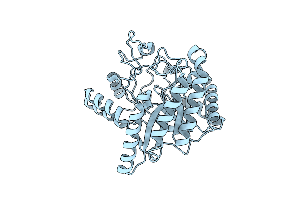
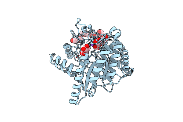
Neutron Structure Of Cellulase Cel6A From Phanerochaete Chrysosporium At Room Temperature
Organism: Phanerodontia chrysosporium
Method: X-RAY DIFFRACTION, NEUTRON DIFFRACTION Resolution:1.36 Å, 1.86 Å Release Date: 2025-03-12 Classification: HYDROLASE |
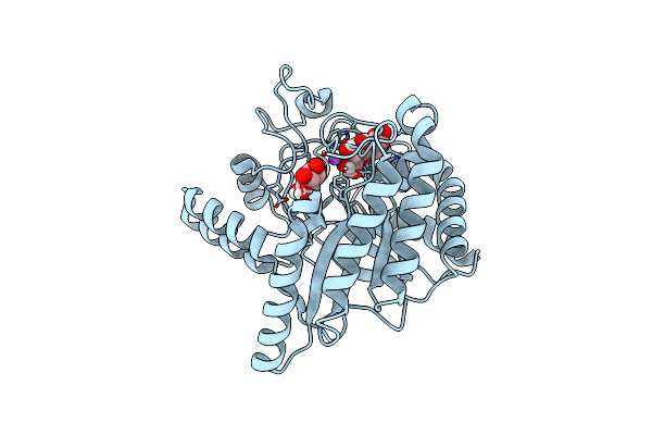 |
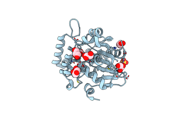
Neutron Structure Of Cellulase Cel6A From Phanerochaete Chrysosporium At Room Temperature, Enzyme-Product Complex
Organism: Phanerodontia chrysosporium
Method: X-RAY DIFFRACTION, NEUTRON DIFFRACTION Resolution:1.40 Å, 1.8 Å Release Date: 2025-03-12 Classification: HYDROLASE Ligands: BGC, NA |
 |
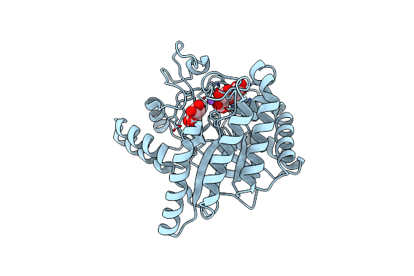
Neutron Structure Of Cellulase Cel6A From Phanerochaete Chrysosporium At Room Temperature, Low-D2O-Solvent
Organism: Phanerodontia chrysosporium
Method: X-RAY DIFFRACTION, NEUTRON DIFFRACTION Resolution:1.40 Å, 2.15 Å Release Date: 2025-03-12 Classification: HYDROLASE |
 |
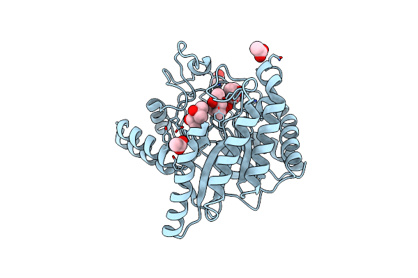
Neutron Structure Of Cellulase Cel6A From Phanerochaete Chrysosporium At Room Temperature, Enzyme-Product Complex, H2O Solvent
Organism: Phanerodontia chrysosporium
Method: X-RAY DIFFRACTION, NEUTRON DIFFRACTION Resolution:1.80 Å, 2.59 Å Release Date: 2025-03-12 Classification: HYDROLASE Ligands: BGC, NA |
 |
High-Resolution X-Ray Structure Of Cellulase Cel6A From Phanerochaete Chrysosporium At Cryogenic Temperature
Organism: Phanerodontia chrysosporium
Method: X-RAY DIFFRACTION Resolution:0.80 Å Release Date: 2025-03-12 Classification: HYDROLASE Ligands: MPD, ACT |
 |
High-Resolution X-Ray Structure Of Cellulase Cel6A From Phanerochaete Chrysosporium At Cryogenic Temperature, Enzyme-Product Complex
Organism: Phanerodontia chrysosporium
Method: X-RAY DIFFRACTION Resolution:0.85 Å Release Date: 2025-03-12 Classification: HYDROLASE Ligands: BGC, NA, CL, PEG |
 |
Organism: Arabidopsis thaliana
Method: X-RAY DIFFRACTION Resolution:2.08 Å Release Date: 2025-02-19 Classification: SIGNALING PROTEIN Ligands: GOL, A1L2E, SO4 |
 |
Organism: Arabidopsis thaliana
Method: X-RAY DIFFRACTION Resolution:1.49 Å Release Date: 2025-02-19 Classification: SIGNALING PROTEIN Ligands: GOL, SO4 |
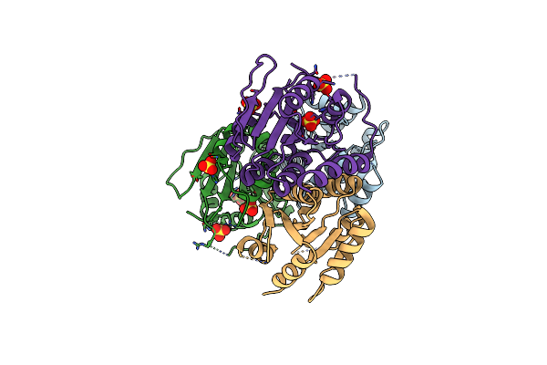 |
Crystal Structure Of Phosphonopyruvate Decarboxylase Rhief From Bacillus Subtilis Atcc6633
Organism: Bacillus spizizenii atcc 6633 = jcm 2499
Method: X-RAY DIFFRACTION Resolution:2.46 Å Release Date: 2024-12-18 Classification: LYASE Ligands: SO4 |
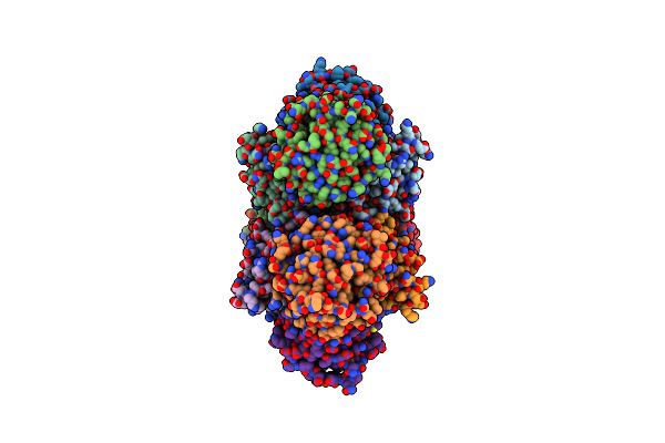 |
Crystal Structure Of Phosphonopyruvate Decarboxylase Rhief From Bacillus Subtilis Atcc6633 In Complex With Thiamine Pyrophosphate
Organism: Bacillus spizizenii atcc 6633 = jcm 2499
Method: X-RAY DIFFRACTION Resolution:3.05 Å Release Date: 2024-12-18 Classification: LYASE Ligands: MG, TPP, SPV |
 |
Organism: Streptomyces mobaraensis
Method: X-RAY DIFFRACTION Resolution:2.00 Å Release Date: 2024-09-18 Classification: TRANSFERASE Ligands: MES, CL, PEG |
 |
Organism: Serratia marcescens
Method: X-RAY DIFFRACTION Resolution:2.20 Å Release Date: 2024-09-04 Classification: HYDROLASE Ligands: GOL |
 |
Crystal Structure Of Pyruvic Oxime Dioxygenase (Pod) From Bradyrhizobium Sp. Wsm3983
Organism: Bradyrhizobium sp. wsm3983
Method: X-RAY DIFFRACTION Resolution:2.47 Å Release Date: 2024-04-03 Classification: OXIDOREDUCTASE Ligands: NI, SO4 |
 |
Crystal Structure Of Pyruvic Oxime Dioxygenase (Pod) From Alcaligenes Faecalis Deleted N-Terminal 18 Residues
Organism: Alcaligenes faecalis subsp. faecalis nbrc 13111
Method: X-RAY DIFFRACTION Resolution:1.55 Å Release Date: 2024-03-20 Classification: OXIDOREDUCTASE Ligands: NI |
 |
Crystal Structure Of Pyruvic Oxime Dioxygenase (Pod) From Alcaligenes Faecalis
Organism: Alcaligenes faecalis subsp. faecalis nbrc 13111
Method: X-RAY DIFFRACTION Resolution:1.77 Å Release Date: 2024-03-06 Classification: OXIDOREDUCTASE Ligands: FE2 |
 |
Organism: Vibrio parahaemolyticus
Method: X-RAY DIFFRACTION Resolution:2.50 Å Release Date: 2023-12-20 Classification: HYDROLASE Ligands: ACT, CA |
 |
Group Deposition Sars-Cov-2 Main Protease In Complex With Inhibitors From The Covid Moonshot -- Crystal Structure Of Sars-Cov-2 Main Protease In Complex With Mat-Pos-F7918075-2 (Sars2_Mproa-X0854)
Organism: Severe acute respiratory syndrome coronavirus 2
Method: X-RAY DIFFRACTION Resolution:1.77 Å Release Date: 2023-11-08 Classification: VIRAL PROTEIN Ligands: DMS, CL, KFU |
 |
Group Deposition Sars-Cov-2 Main Protease In Complex With Inhibitors From The Covid Moonshot -- Crystal Structure Of Sars-Cov-2 Main Protease In Complex With Mat-Pos-E194Df51-1 (Sars2_Mproa-X0862)
Organism: Severe acute respiratory syndrome coronavirus 2
Method: X-RAY DIFFRACTION Resolution:1.81 Å Release Date: 2023-11-08 Classification: VIRAL PROTEIN Ligands: DMS, CL, KG9 |
 |
Group Deposition Sars-Cov-2 Main Protease In Complex With Inhibitors From The Covid Moonshot -- Crystal Structure Of Sars-Cov-2 Main Protease In Complex With Gab-Rev-70Cc3Ca5-4 (Mpro-X10019)
Organism: Severe acute respiratory syndrome coronavirus 2
Method: X-RAY DIFFRACTION Resolution:1.71 Å Release Date: 2023-11-08 Classification: VIRAL PROTEIN Ligands: KJI, DMS |
 |
Group Deposition Sars-Cov-2 Main Protease In Complex With Inhibitors From The Covid Moonshot -- Crystal Structure Of Sars-Cov-2 Main Protease In Complex With Ben-Dnd-031A96Cc-8 (Mpro-X10022)
Organism: Severe acute respiratory syndrome coronavirus 2
Method: X-RAY DIFFRACTION Resolution:1.30 Å Release Date: 2023-11-08 Classification: VIRAL PROTEIN Ligands: KJO, DMS |