
Search Count: 20
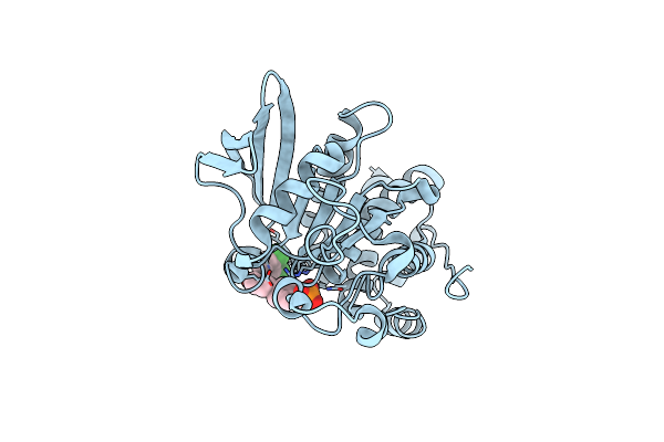 |
X-Ray Characterization Of Striatal-Enriched Protein Tyrosine Phosphatase Inhibitors
Organism: Homo sapiens
Method: X-RAY DIFFRACTION Resolution:2.15 Å Release Date: 2017-11-22 Classification: HYDROLASE Ligands: AXK |
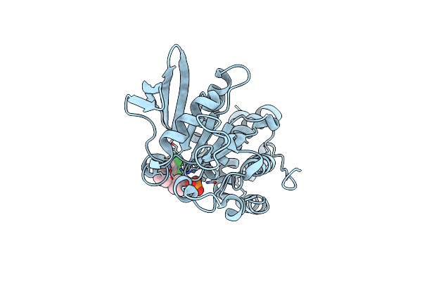 |
X-Ray Characterization Of Striatal-Enriched Protein Tyrosine Phosphatase Inhibitors
Organism: Homo sapiens
Method: X-RAY DIFFRACTION Resolution:2.10 Å Release Date: 2017-11-22 Classification: HYDROLASE Ligands: AY5 |
 |
X-Ray Characterization Of Striatal-Enriched Protein Tyrosine Phosphatase Inhibitors
Organism: Homo sapiens
Method: X-RAY DIFFRACTION Resolution:2.05 Å Release Date: 2017-11-22 Classification: HYDROLASE Ligands: AY8 |
 |
Organism: Homo sapiens
Method: X-RAY DIFFRACTION Resolution:1.45 Å Release Date: 2014-03-26 Classification: HYDROLASE Ligands: MN, PO4, CL |
 |
Organism: Homo sapiens, Rattus norvegicus
Method: X-RAY DIFFRACTION Resolution:2.20 Å Release Date: 2014-03-26 Classification: HYDROLASE/NUCLEAR PROTEIN Ligands: MN, CL, PO4, GOL |
 |
Organism: Homo sapiens, Rattus norvegicus
Method: X-RAY DIFFRACTION Resolution:2.10 Å Release Date: 2014-03-26 Classification: HYDROLASE Ligands: MN, GOL, PO4 |
 |
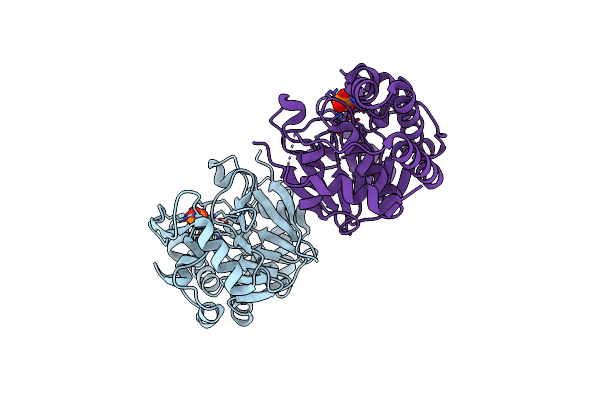
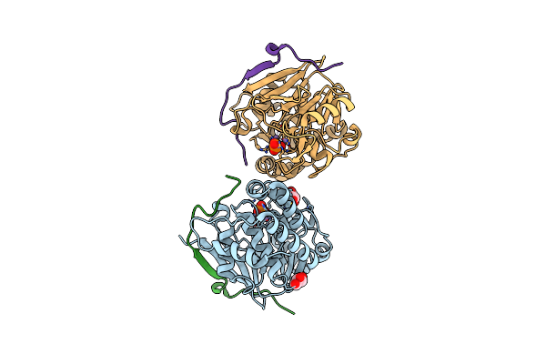
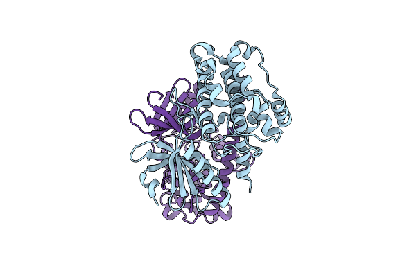
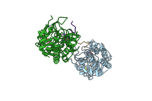
Crystal Structure Of A Complex Between Protein Phosphatase 1 Alpha (Pp1) And The Pp1 Binding And Pdz Domains Of Spinophilin
Organism: Homo sapiens, Rattus norvegicus
Method: X-RAY DIFFRACTION Resolution:1.85 Å Release Date: 2010-03-23 Classification: HYDROLASE Ligands: GOL, MN, MES |
 |
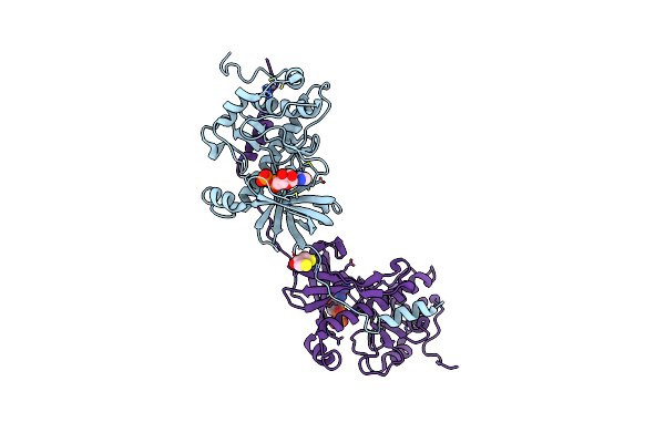
Crystal Structure Of A Complex Between Protein Phosphatase 1 Alpha (Pp1), The Pp1 Binding And Pdz Domains Of Spinophilin And The Small Natural Molecular Toxin Nodularin-R
Organism: Homo sapiens, Rattus norvegicus, Nodularia spumigena
Method: X-RAY DIFFRACTION Resolution:2.00 Å Release Date: 2010-03-23 Classification: HYDROLASE/HYDROLASE INHIBITOR Ligands: GOL, MN |
 |
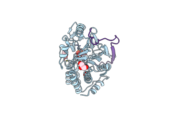
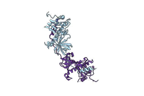
Crystal Structure Of A Complex Between Protein Phosphatase 1 Alpha (Pp1) And The Pp1 Binding And Pdz Domains Of Neurabin
Organism: Homo sapiens, Rattus norvegicus
Method: X-RAY DIFFRACTION Resolution:2.20 Å Release Date: 2010-03-23 Classification: HYDROLASE/HYDROLASE REGULATOR Ligands: MN, PO4, GOL |
 |
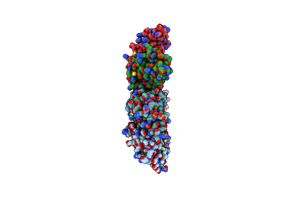
Organism: Rattus norvegicus
Method: SOLUTION NMR Release Date: 2007-03-13 Classification: PROTEIN BINDING |
 |
Organism: Rattus norvegicus
Method: SOLUTION NMR Release Date: 2007-01-09 Classification: SIGNALING PROTEIN |
 |
Organism: Rattus norvegicus
Method: SOLUTION NMR Release Date: 2007-01-09 Classification: PROTEIN BINDING |
 |
Organism: Caenorhabditis elegans
Method: X-RAY DIFFRACTION Resolution:2.64 Å Release Date: 2006-02-07 Classification: TRANSFERASE |
 |
Crystal Structure Of The Auto-Inhibited Kinase Domain Of Calcium/Calmodulin Activated Kinase Ii
Organism: Caenorhabditis elegans
Method: X-RAY DIFFRACTION Resolution:1.80 Å Release Date: 2005-12-13 Classification: TRANSFERASE |
 |
Organism: Mus musculus
Method: X-RAY DIFFRACTION Resolution:2.65 Å Release Date: 2003-06-02 Classification: TRANSFERASE Ligands: DTT, CL, TBR |
 |
Crystal Structure Of The Atypical Protein Kinase Domain Of A Trp Ca-Channel, Chak (Amppnp Complex)
Organism: Mus musculus
Method: X-RAY DIFFRACTION Resolution:2.00 Å Release Date: 2001-06-06 Classification: TRANSFERASE Ligands: ZN, ANP, DTT |
 |
Crystal Structure Of The Atypical Protein Kinase Domain Of A Trp Ca-Channel, Chak (Adp-Mg Complex)
Organism: Mus musculus
Method: X-RAY DIFFRACTION Resolution:2.40 Å Release Date: 2001-06-06 Classification: TRANSFERASE Ligands: ZN, ADP, MG, DTT |
 |
Crystal Structure Of The Atypical Protein Kinase Domain Of A Trp Ca-Channel, Chak (Apo)
Organism: Mus musculus
Method: X-RAY DIFFRACTION Resolution:2.80 Å Release Date: 2001-06-06 Classification: TRANSFERASE Ligands: ZN |
 |
Organism: Rattus norvegicus
Method: X-RAY DIFFRACTION Resolution:2.50 Å Release Date: 1998-04-08 Classification: KINASE |
 |
Protein Serine/Threonine Phosphatase-1 (Alpha Isoform, Type 1) Complexed With Microcystin-Lr Toxin
Organism: Oryctolagus cuniculus, Microcystis aeruginosa
Method: X-RAY DIFFRACTION Resolution:2.10 Å Release Date: 1996-06-20 Classification: HYDROLASE/HYDROLASE INHIBITOR Ligands: MN, BME |