
Search Count: 25
 |
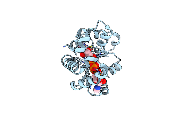
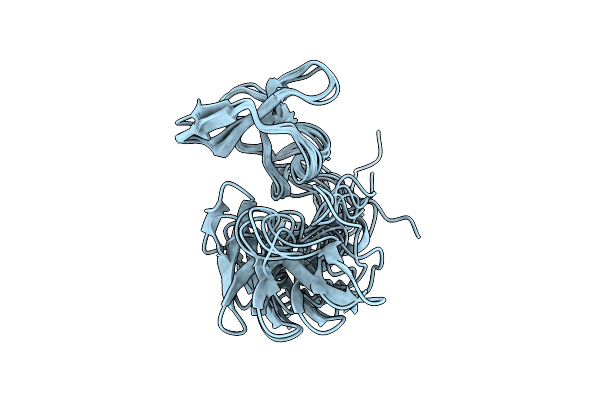
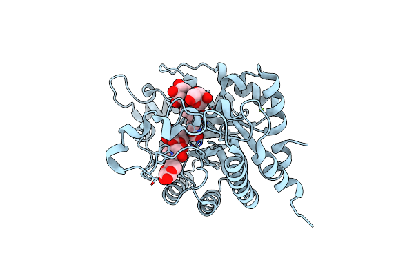
Panoptes Opts Minimal Crispr Polymerase (Mcpol) From Klebsiella Pneumoniae Strain Kp67
Organism: Klebsiella pneumoniae
Method: X-RAY DIFFRACTION Release Date: 2025-09-24 Classification: ANTIVIRAL PROTEIN |
 |
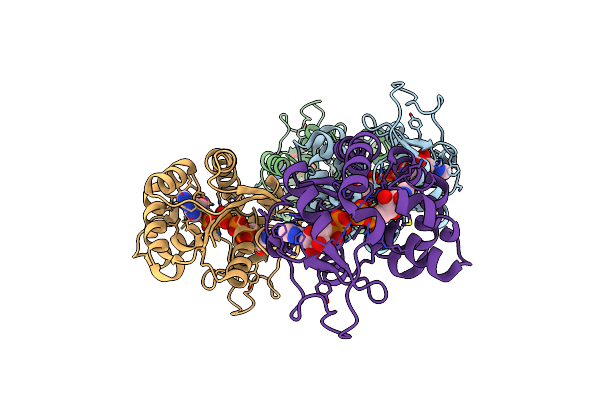
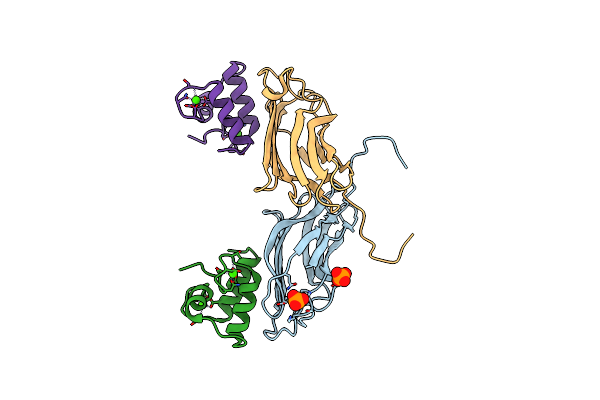
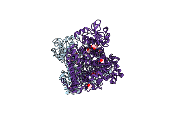
Panoptes Opts Minimal Crispr Polymerase (Mcpol) With Non-Hydrolyzable Ligand Apcpp From Klebsiella Pneumoniae Strain Kp67
Organism: Klebsiella pneumoniae
Method: X-RAY DIFFRACTION Release Date: 2025-09-24 Classification: ANTIVIRAL PROTEIN Ligands: APC, MG |
 |
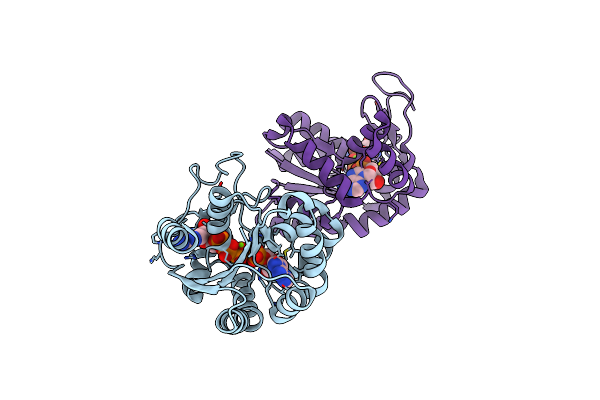
Organism: Escherichia coli k-12
Method: X-RAY DIFFRACTION Resolution:2.00 Å Release Date: 2025-02-05 Classification: SIGNALING PROTEIN Ligands: AP5 |
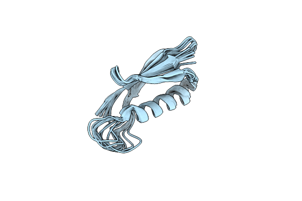 |
Organism: Escherichia coli
Method: SOLUTION NMR Release Date: 2024-10-02 Classification: PROTEIN BINDING |
 |
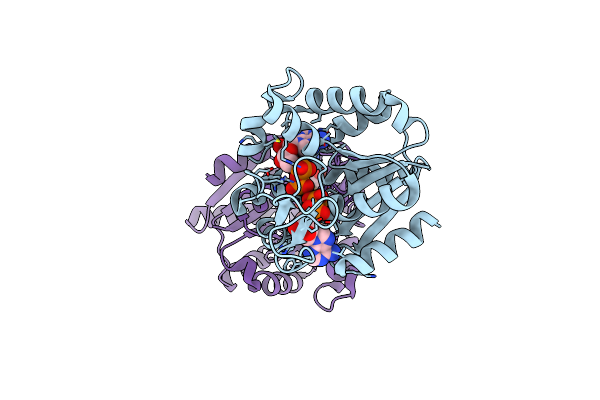
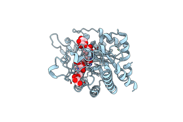
E. Coli Adenylate Kinase In Complex With Two Adp Molecules And Mg2+ As A Result Of Enzymatic Ap4A Hydrolysis
Organism: Escherichia coli
Method: X-RAY DIFFRACTION Resolution:2.11 Å Release Date: 2024-07-10 Classification: TRANSFERASE Ligands: ADP, MG, CL |
 |
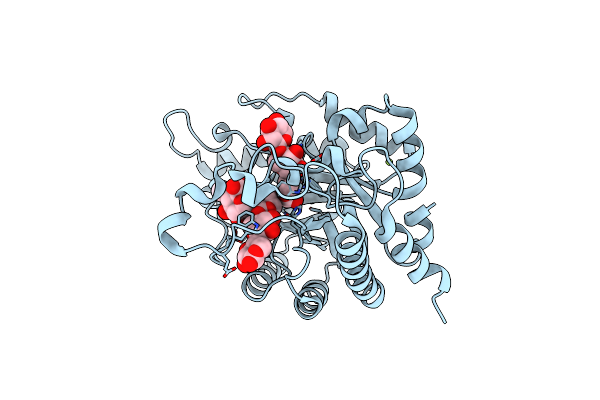
E. Coli Adenylate Kinase In Complex With Atp And Amp And Mg2+ As A Result Of Enzymatic Ap4A Hydrolysis.
Organism: Escherichia coli
Method: X-RAY DIFFRACTION Resolution:1.90 Å Release Date: 2024-07-10 Classification: TRANSFERASE Ligands: ATP, ADP, AMP, MG |
 |
E. Coli Adenylate Kinase Asp84Ala Variant In Complex With Two Adp Molecules As A Result Of Enzymatic Ap4A Hydrolysis.
Organism: Escherichia coli
Method: X-RAY DIFFRACTION Resolution:1.59 Å Release Date: 2024-07-10 Classification: TRANSFERASE Ligands: ADP |
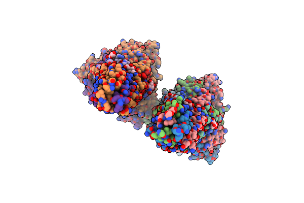 |
Crystal Structure Of A Human Metapneumovirus Subtype B2 Trimeric Fusion Protein
Organism: Human metapneumovirus
Method: X-RAY DIFFRACTION Resolution:2.81 Å Release Date: 2021-06-30 Classification: VIRAL PROTEIN Ligands: NAG |
 |
Differential Regulation Of The Xylan Degrading Apparatus Of Cellvibrio Japonicus By A Novel Two Component System
Organism: Cellvibrio japonicus
Method: X-RAY DIFFRACTION Resolution:2.60 Å Release Date: 2008-10-14 Classification: TRANSFERASE Ligands: PO4, CL |
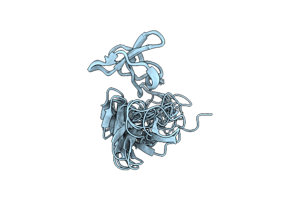 |
Organism: Piromyces equi
Method: SOLUTION NMR Release Date: 2007-09-25 Classification: PROTEIN BINDING |
 |
Organism: Piromyces equi
Method: SOLUTION NMR Release Date: 2007-09-25 Classification: PROTEIN BINDING |
 |
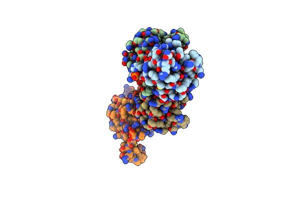
The S45A, T46A Mutant Of The Type I Cohesin-Dockerin Complex From The Cellulosome Of Clostridium Thermocellum
Organism: Clostridium thermocellum
Method: X-RAY DIFFRACTION Resolution:2.03 Å Release Date: 2007-02-13 Classification: CELL ADHESION Ligands: PO4, CA |
 |
Xylanase Xyn10B Mutant (E262S) From Cellvibrio Mixtus In Complex With Xylopentaose
Organism: Cellvibrio mixtus
Method: X-RAY DIFFRACTION Resolution:1.72 Å Release Date: 2003-12-18 Classification: HYDROLASE Ligands: MG |
 |
Xylanase Xyn10B Mutant (E262S) From Cellvibrio Mixtus In Complex With 4-O-Methyl Glucuronic Acid
Organism: Cellvibrio mixtus
Method: X-RAY DIFFRACTION Resolution:1.55 Å Release Date: 2003-12-18 Classification: HYDROLASE Ligands: CL, MG |
 |
Xylanase Xyn10B Mutant (E262S) From Cellvibrio Mixtus In Complex With Arabinofuranose Alpha-1,3 Linked To Xylobiose
Organism: Cellvibrio mixtus
Method: X-RAY DIFFRACTION Resolution:1.43 Å Release Date: 2003-12-18 Classification: HYDROLASE Ligands: CL, MG |
 |
Xylanase Xyn10B Mutant (E262S) From Cellvibrio Mixtus In Complex With Arabinofuranose Alpha 1,3 Linked To Xylotriose
Organism: Cellvibrio mixtus
Method: X-RAY DIFFRACTION Resolution:1.60 Å Release Date: 2003-12-18 Classification: HYDROLASE Ligands: CL, MG |
 |
Pseudomonas Cellulosa E292A Alpha-D-Glucuronidase Mutant Complexed With Aldotriuronic Acid
Organism: Pseudomonas cellulosa
Method: X-RAY DIFFRACTION Resolution:1.50 Å Release Date: 2003-05-01 Classification: HYDROLASE Ligands: GCV, EDO, CO |
 |
Organism: Pseudomonas cellulosa
Method: X-RAY DIFFRACTION Resolution:1.48 Å Release Date: 2002-09-26 Classification: GLUCURONIDASE Ligands: EDO, CO, MG |
 |
Structure Of Pseudomonas Cellulosa Alpha-D-Glucuronidase Complexed With Xylobiose
Organism: Pseudomonas cellulosa
Method: X-RAY DIFFRACTION Resolution:1.90 Å Release Date: 2002-09-26 Classification: GLUCURONIDASE Ligands: EDO, CO |
 |
Structure Of Pseudomonas Cellulosa Alpha-D-Glucuronidase Complexed With Glucuronic Acid
Organism: Cellvibrio japonicus
Method: X-RAY DIFFRACTION Resolution:1.90 Å Release Date: 2002-09-26 Classification: HYDROLASE Ligands: BDP, EDO, CO |