
Search Count: 29
 |
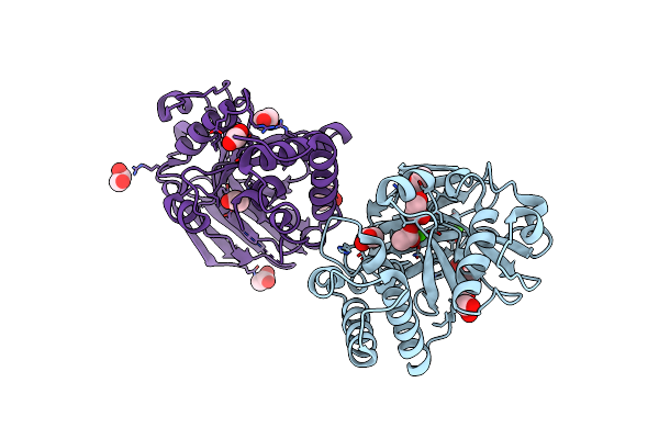
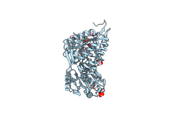
Structure And Mechanism Of Salicylate Hydroxylase From Pseudomonas Putida G7
Organism: Pseudomonas putida
Method: X-RAY DIFFRACTION Resolution:2.01 Å Release Date: 2018-12-26 Classification: OXIDOREDUCTASE Ligands: IOD, GOL, EDO, SO4 |
 |
Crystal Structure Of An Antigenic Nucleotidyltransferase-Like Protein From Paracoccidioides Brasiliensis
Organism: Paracoccidioides brasiliensis (strain pb18)
Method: X-RAY DIFFRACTION Resolution:1.86 Å Release Date: 2018-01-31 Classification: TRANSFERASE Ligands: MG |
 |
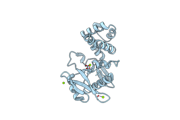
Organism: Pseudomonas putida
Method: X-RAY DIFFRACTION Resolution:1.74 Å Release Date: 2016-05-04 Classification: LYASE Ligands: EDO, CL, ACT |
 |
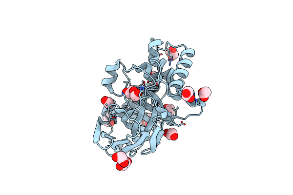
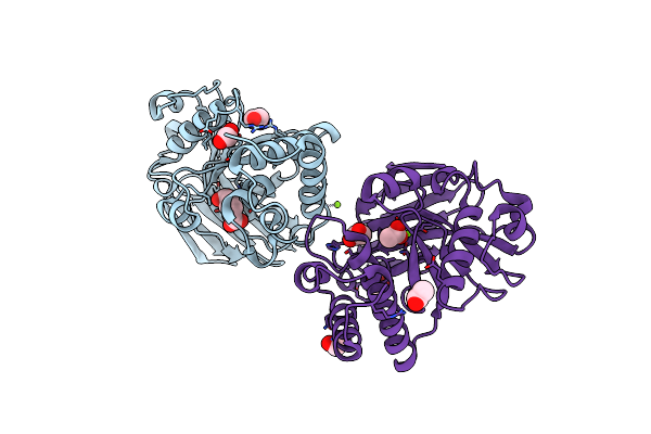
4-Oxalocrotonate Decarboxylase From Pseudomonas Putida G7 - Complexed With Magnesium
Organism: Pseudomonas putida
Method: X-RAY DIFFRACTION Resolution:1.90 Å Release Date: 2016-05-04 Classification: LYASE Ligands: MG, EDO, CL, SO4, ACT |
 |
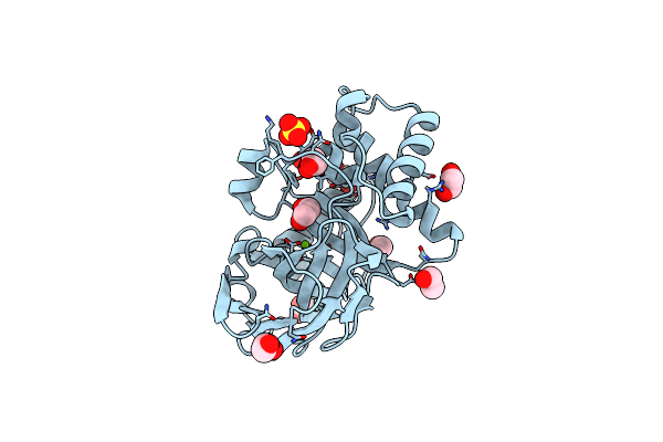
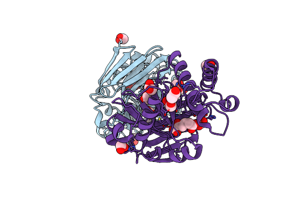
4-Oxalocrotonate Decarboxylase From Pseudomonas Putida G7 - Complexed With Magnesium And Alpha-Ketoglutarate
Organism: Pseudomonas putida
Method: X-RAY DIFFRACTION Resolution:1.94 Å Release Date: 2016-05-04 Classification: LYASE Ligands: MG, AKG, EDO, CL, ACT, SO4 |
 |
4-Oxalocrotonate Decarboxylase From Pseudomonas Putida G7 - Complexed With Calcium And Acetate
Organism: Pseudomonas putida
Method: X-RAY DIFFRACTION Resolution:1.78 Å Release Date: 2016-05-04 Classification: LYASE Ligands: CA, EDO, ACT |
 |
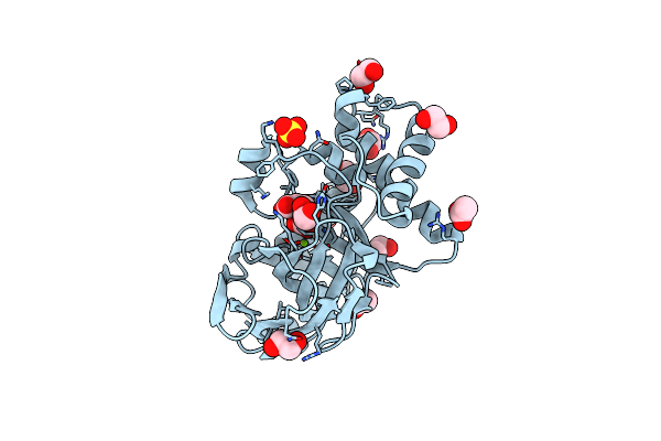
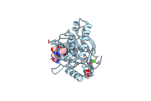
4-Oxalocrotonate Decarboxylase From Pseudomonas Putida G7 - Complexed With Magnesium And Adipate
Organism: Pseudomonas putida
Method: X-RAY DIFFRACTION Resolution:1.72 Å Release Date: 2016-05-04 Classification: LYASE Ligands: MG, 0L1, EDO, ACT |
 |
4-Oxalocrotonate Decarboxylase From Pseudomonas Putida G7 - Complexed With Magnesium And 2-Oxoadipate
Organism: Pseudomonas putida
Method: X-RAY DIFFRACTION Resolution:1.57 Å Release Date: 2016-05-04 Classification: LYASE Ligands: MG, OOG, EDO, ACT |
 |
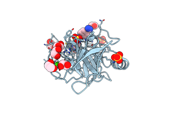
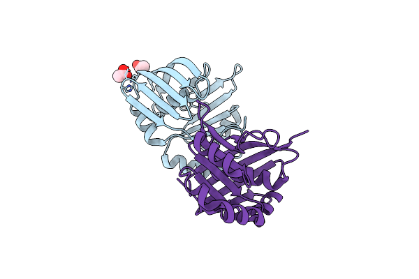
Crystal Structure Of Leucurolysin-A Complexed With An Endogenous Tripeptide (Qsw).
Organism: Bothrops leucurus
Method: X-RAY DIFFRACTION Resolution:1.90 Å Release Date: 2015-04-08 Classification: HYDROLASE Ligands: CA, ZN, ACT, EDO |
 |
Crystal Structure Of A Salicylaldehyde Dehydrogenase From Pseudomonas Putida G7 Complexed With Salicylaldehyde
Organism: Pseudomonas putida
Method: X-RAY DIFFRACTION Resolution:2.42 Å Release Date: 2014-04-02 Classification: OXIDOREDUCTASE Ligands: NK, SO4, EDO |
 |
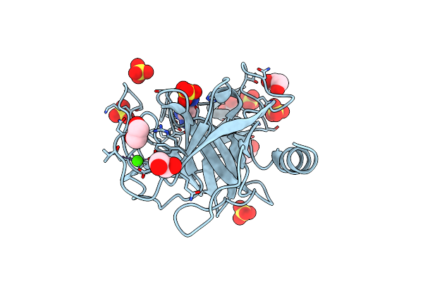
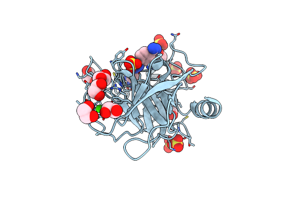
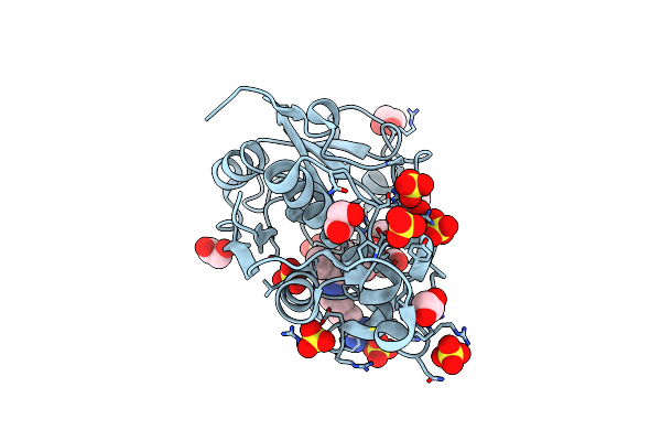
A Comparative Study On The Inhibition Of Bovine Beta-Trypsin By Bis-Benzamidines Diminazene And Pentamidine By X-Ray Crystallography And Itc
Organism: Bos taurus
Method: X-RAY DIFFRACTION Resolution:1.57 Å Release Date: 2010-03-23 Classification: HYDROLASE Ligands: CA, BRN, EDO, SO4 |
 |
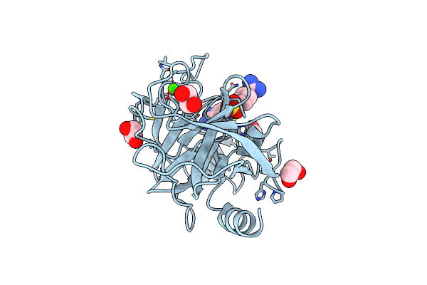
A Comparative Study On The Inhibition Of Bovine Beta-Trypsin By Bis-Benzamidines Diminazene And Pentamidine By X-Ray Crystallography And Itc
Organism: Bos taurus
Method: X-RAY DIFFRACTION Resolution:1.70 Å Release Date: 2010-03-23 Classification: HYDROLASE Ligands: CA, PNT, EDO |
 |
A Comparative Study On The Inhibition Of Bovine Beta-Trypsin By Bis-Benzamidines Diminazene And Pentamidine By X-Ray Crystallography And Itc
Organism: Bos taurus
Method: X-RAY DIFFRACTION Resolution:1.55 Å Release Date: 2010-03-23 Classification: HYDROLASE Ligands: CA, PBZ, EDO, SO4 |
 |
A Comparative Study On The Inhibition Of Bovine Beta-Trypsin By Bis-Benzamidines Diminazene And Pentamidine By X-Ray Crystallography And Itc
Organism: Bos taurus
Method: X-RAY DIFFRACTION Resolution:1.57 Å Release Date: 2010-03-23 Classification: HYDROLASE Ligands: CA, BRN, EDO, SO4 |
 |
A Comparative Study On The Inhibition Of Bovine Beta-Trypsin By The Bis-Benzamidines Diminazene And Pentamidine
Organism: Bos taurus
Method: X-RAY DIFFRACTION Resolution:1.70 Å Release Date: 2010-03-23 Classification: HYDROLASE Ligands: CA, BRN, EDO, SO4 |
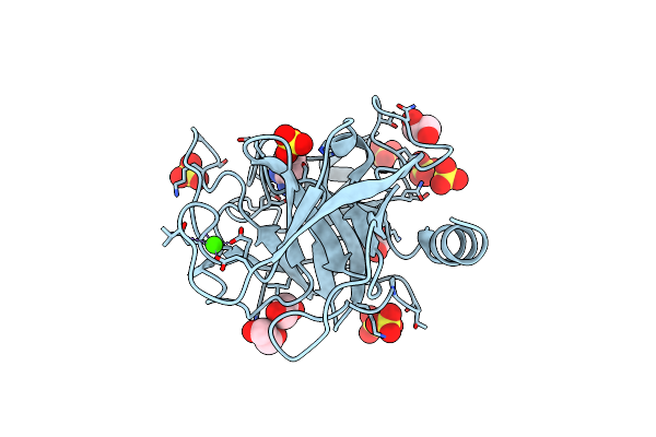 |
A Comparative Study On The Inhibition Of Bovine Beta-Trypsin By Bis-Benzamidines Diminazene And Pentamidine By X-Ray Crystallography And Itc
Organism: Bos taurus
Method: X-RAY DIFFRACTION Resolution:1.55 Å Release Date: 2010-03-23 Classification: HYDROLASE Ligands: CA, BEN, EDO, SO4 |
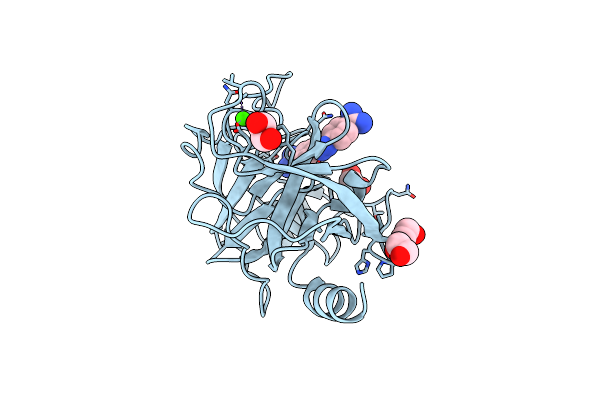 |
A Comparative Study On The Inhibition Of Bovine Beta-Trypsin By Bis-Benzamidines Diminazene And Pentamidine By X-Ray Crystallography And Itc
Organism: Bos taurus
Method: X-RAY DIFFRACTION Resolution:1.75 Å Release Date: 2010-03-23 Classification: HYDROLASE Ligands: CA, BRN, EDO |
 |
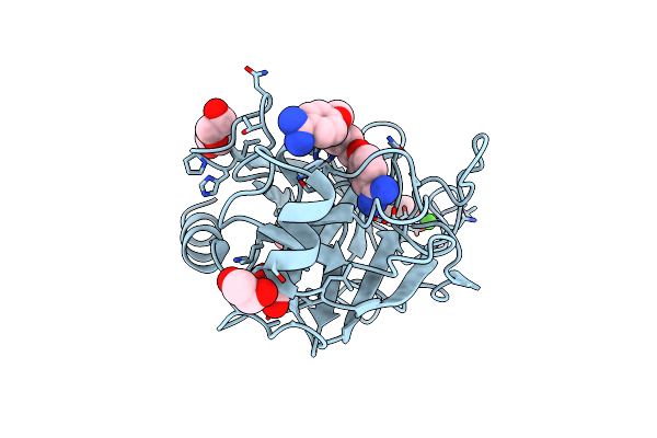
Crystal Structure Of The Carica Candamarcensis Cysteine Protease Cms1Ms2 In Complex With E-64.
Organism: Carica candamarcensis
Method: X-RAY DIFFRACTION Resolution:1.87 Å Release Date: 2010-02-16 Classification: HYDROLASE Ligands: E64, SO4, EDO |
 |
The Crystal Structure Of Yaeq Protein From Xanthomonas Axonopodis Pv. Citri
Organism: Xanthomonas axonopodis pv. citri
Method: X-RAY DIFFRACTION Resolution:1.90 Å Release Date: 2007-02-27 Classification: UNKNOWN FUNCTION Ligands: ACT |
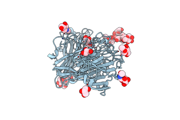 |
Crystal Structure Of Exo-Inulinase From Aspergillus Awamori In Spacegroup P212121
Organism: Aspergillus awamori
Method: X-RAY DIFFRACTION Resolution:1.89 Å Release Date: 2004-12-28 Classification: HYDROLASE Ligands: MAN, NAG, GOL |