
Search Count: 55
 |
Structure Of 6-Aminohexanoate-Oligomer Hydrolase Nylc, D122G/H130Y/T267C Mutant, Hydroxylamine-Treated
Organism: Arthrobacter
Method: X-RAY DIFFRACTION Resolution:1.21 Å Release Date: 2023-03-29 Classification: HYDROLASE Ligands: GOL, SO4 |
 |
Structure Of 6-Aminohexanoate-Oligomer Hydrolase Nylc Precursor, H130Y/N266A/T267A Mutant
Organism: Arthrobacter
Method: X-RAY DIFFRACTION Resolution:1.35 Å Release Date: 2023-03-01 Classification: HYDROLASE Ligands: GOL, SO4, NA |
 |
Structure Of 6-Aminohexanoate-Oligomer Hydrolase Nylc Precursor, D122G/H130Y/T267C Mutant
Organism: Arthrobacter
Method: X-RAY DIFFRACTION Resolution:1.13 Å Release Date: 2023-03-01 Classification: HYDROLASE Ligands: GOL, NA, SO4 |
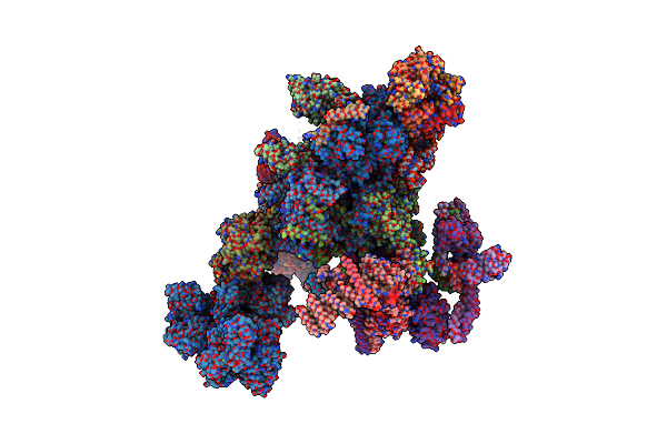 |
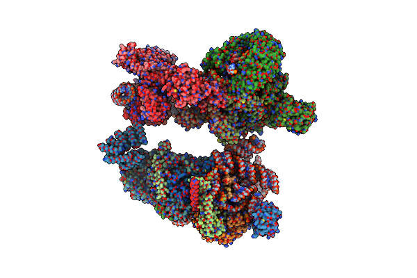
Yeast C Complex Spliceosome At 2.8 Angstrom Resolution With Prp18/Slu7 Bound
Organism: Saccharomyces cerevisiae
Method: ELECTRON MICROSCOPY Release Date: 2021-03-10 Classification: SPLICING Ligands: MG, K, KGN, GTP, ZN |
 |
Organism: Homo sapiens
Method: ELECTRON MICROSCOPY Release Date: 2019-04-17 Classification: SPLICING Ligands: IHP, MG, GTP, ZN |
 |
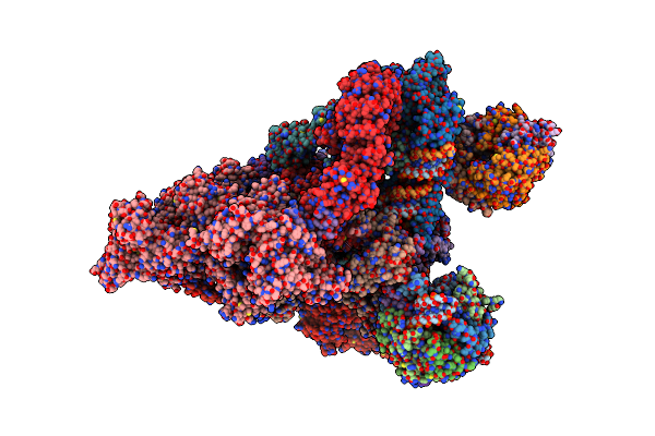
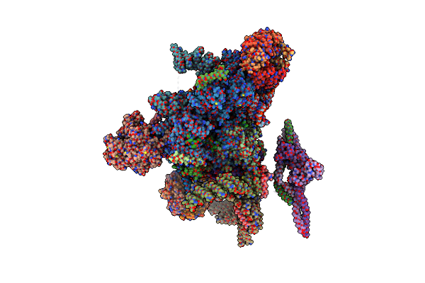
Structure Of A Human Fully-Assembled Precatalytic Spliceosome (Pre-B Complex).
Organism: Homo sapiens, Human adenovirus 2
Method: ELECTRON MICROSCOPY Release Date: 2019-04-17 Classification: SPLICING Ligands: ZN, MG, GTP, IHP |
 |
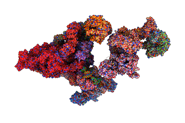
Organism: Homo sapiens, Human adenovirus 2
Method: ELECTRON MICROSCOPY Release Date: 2019-02-20 Classification: SPLICING Ligands: MG, K, ATP, GTP, ZN, IHP |
 |
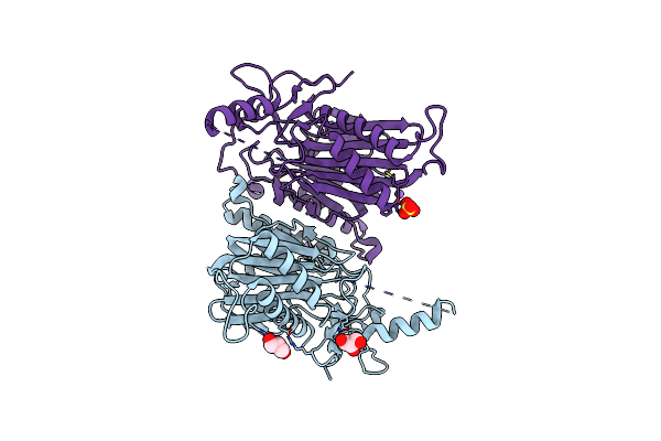
Structure Of 6-Aminohexanoate-Oligomer Hydrolase From Arthrobacter Sp. Ki72.
Organism: Arthrobacter sp.
Method: X-RAY DIFFRACTION Resolution:1.60 Å Release Date: 2018-11-21 Classification: HYDROLASE Ligands: GOL, SO4, CL |
 |
Structure Of 6-Aminohexanoate-Oligomer Hydrolase From Arthrobacter Sp. Ki72., D122G Mutant
Organism: Flavobacterium sp. ki723t1
Method: X-RAY DIFFRACTION Resolution:2.00 Å Release Date: 2018-11-21 Classification: HYDROLASE Ligands: SO4, CL, GOL |
 |
Structure Of 6-Aminohexanoate-Oligomer Hydrolase From Arthrobacter Sp. Ki72., D122R Mutant
Organism: Flavobacterium sp. ki723t1
Method: X-RAY DIFFRACTION Resolution:1.20 Å Release Date: 2018-11-21 Classification: HYDROLASE Ligands: PO4, GOL |
 |
Structure Of 6-Aminohexanoate-Oligomer Hydrolase From Arthrobacter Sp. Ki72., D122K Mutant
Organism: Flavobacterium sp. ki723t1
Method: X-RAY DIFFRACTION Resolution:1.10 Å Release Date: 2018-11-21 Classification: HYDROLASE Ligands: PO4, GOL |
 |
Structure Of 6-Aminohexanoate-Oligomer Hydrolase From Arthrobacter Sp. Ki72., D122V Mutant
Organism: Flavobacterium sp. ki723t1
Method: X-RAY DIFFRACTION Resolution:1.05 Å Release Date: 2018-11-21 Classification: HYDROLASE Ligands: GOL, PO4 |
 |
Structure Of 6-Aminohexanoate-Oligomer Hydrolase From Arthrobacter Sp. Ki72., H130Y Mutant
Organism: Flavobacterium sp. ki723t1
Method: X-RAY DIFFRACTION Resolution:1.90 Å Release Date: 2018-11-21 Classification: HYDROLASE Ligands: SO4, GOL |
 |
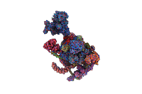
Prespliceosome Structure Provides Insight Into Spliceosome Assembly And Regulation (Map A2)
Organism: Saccharomyces cerevisiae
Method: ELECTRON MICROSCOPY Release Date: 2018-08-22 Classification: SPLICING Ligands: ZN |
 |
Structure Of 6-Aminohexanoate-Oligomer Hydrolase From Arthrobacter Sp. Ki72., D122G/H130Y Mutant
Organism: Flavobacterium sp. ki723t1
Method: X-RAY DIFFRACTION Resolution:1.39 Å Release Date: 2018-07-25 Classification: HYDROLASE Ligands: GOL, NA, SO4 |
 |
Structure Of 6-Aminohexanoate-Oligomer Hydrolase From Arthrobacter Sp. Ki72., D36A/D122G/H130Y/E263Q Mutant
Organism: Flavobacterium sp. ki723t1
Method: X-RAY DIFFRACTION Resolution:1.03 Å Release Date: 2018-07-25 Classification: HYDROLASE Ligands: GOL, PO4, CL |
 |
Organism: Saccharomyces cerevisiae s288c, Saccharomyces cerevisiae (strain atcc 204508 / s288c), Saccharomyces cerevisiae
Method: ELECTRON MICROSCOPY Release Date: 2018-01-17 Classification: SPLICING Ligands: MG, IHP, GTP, ZN |
 |
Organism: Saccharomyces cerevisiae, Saccharomyces cerevisiae , Saccharomyces cerevisiae (strain atcc 204508 / s288c)
Method: ELECTRON MICROSCOPY Release Date: 2017-05-31 Classification: SPLICING Ligands: GTP, ZN |
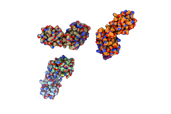 |
Organism: Saccharomyces cerevisiae (strain atcc 204508 / s288c)
Method: X-RAY DIFFRACTION Resolution:2.70 Å Release Date: 2017-04-12 Classification: RNA binding domain |
 |
Organism: Saccharomyces cerevisiae
Method: X-RAY DIFFRACTION Resolution:1.65 Å Release Date: 2017-04-12 Classification: RNA binding domain |