
Search Count: 16
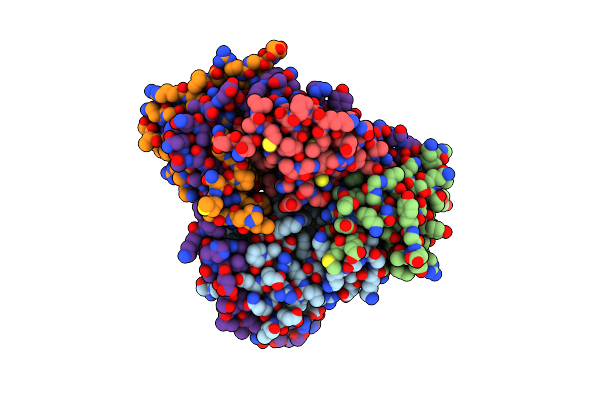 |
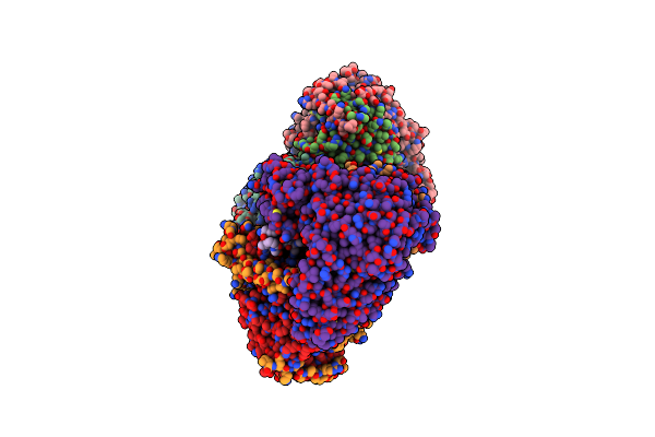
Organism: Homo sapiens
Method: X-RAY DIFFRACTION Resolution:2.40 Å Release Date: 2017-12-06 Classification: PROTEIN BINDING |
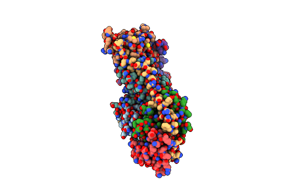 |
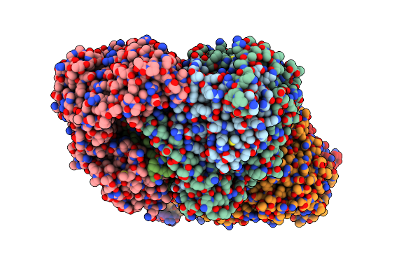
Organism: Homo sapiens, Mus musculus
Method: X-RAY DIFFRACTION Resolution:2.02 Å Release Date: 2017-12-06 Classification: PROTEIN BINDING |
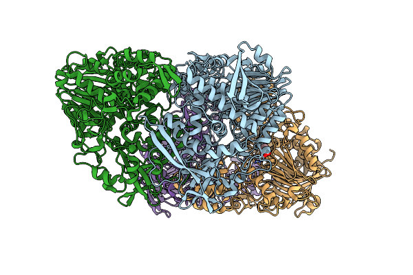 |
Structure Of N-Acylhomoserine Lactone Acylase Macq Shortened Spacer Mutant (Delta202-208) In Uncleaved Form
Organism: Acidovorax sp. mr-s7
Method: X-RAY DIFFRACTION Resolution:1.80 Å Release Date: 2016-06-29 Classification: HYDROLASE Ligands: GOL |
 |
Organism: Acidovorax sp. mr-s7
Method: X-RAY DIFFRACTION Resolution:2.60 Å Release Date: 2016-03-02 Classification: HYDROLASE |
 |
Structure Of N-Acylhomoserine Lactone Acylase Macq In Complex With Decanoic Acid
Organism: Acidovorax sp. mr-s7
Method: X-RAY DIFFRACTION Resolution:2.20 Å Release Date: 2016-03-02 Classification: HYDROLASE Ligands: DKA |
 |
Structure Of N-Acylhomoserine Lactone Acylase Macq In Complex With Phenylacetic Acid
Organism: Acidovorax sp. mr-s7
Method: X-RAY DIFFRACTION Resolution:1.75 Å Release Date: 2016-03-02 Classification: HYDROLASE Ligands: PAC, GOL |
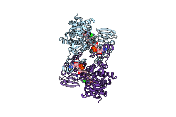 |
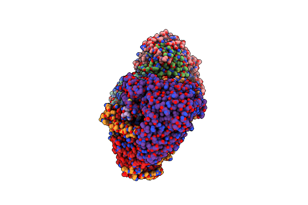
Organism: Homo sapiens
Method: X-RAY DIFFRACTION Resolution:2.70 Å Release Date: 2015-11-25 Classification: HYDROLASE Ligands: NAI, XFA |
 |
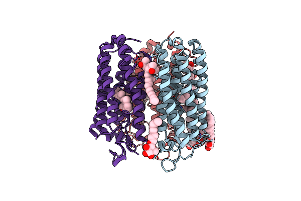
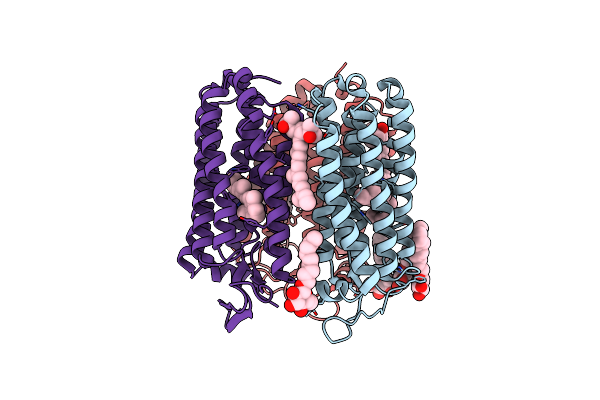
Organism: Halobacterium
Method: X-RAY DIFFRACTION Resolution:1.80 Å Release Date: 2014-10-15 Classification: TRANSPORT PROTEIN Ligands: RET, 22B, SO4, L2P, L3P, L4P, SQL |
 |
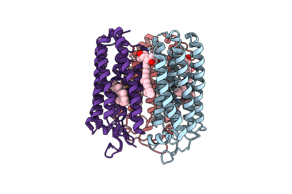
An M-Like Reaction State Of The Azide-Bound Purple Form Of Pharaonis Halorhodopsin
Organism: Natronomonas pharaonis
Method: X-RAY DIFFRACTION Resolution:2.30 Å Release Date: 2013-06-19 Classification: MEMBRANE PROTEIN Ligands: RET, BNG, 22B, L3P, AZI |
 |
Organism: Natronomonas pharaonis
Method: X-RAY DIFFRACTION Resolution:1.80 Å Release Date: 2011-08-31 Classification: MEMBRANE PROTEIN Ligands: RET, BNG, 22B |
 |
Organism: Natronomonas pharaonis
Method: X-RAY DIFFRACTION Resolution:2.10 Å Release Date: 2011-08-31 Classification: MEMBRANE PROTEIN Ligands: RET, BNG, 22B |
 |
Organism: Natronomonas pharaonis
Method: X-RAY DIFFRACTION Resolution:2.20 Å Release Date: 2011-08-31 Classification: MEMBRANE PROTEIN Ligands: RET, 22B, BR |
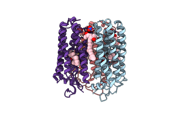 |
Organism: Natronomonas pharaonis
Method: X-RAY DIFFRACTION Resolution:2.20 Å Release Date: 2011-08-31 Classification: MEMBRANE PROTEIN Ligands: RET, NO3, 22B |
 |
Organism: Natronomonas pharaonis
Method: X-RAY DIFFRACTION Resolution:1.90 Å Release Date: 2011-02-02 Classification: MEMBRANE PROTEIN Ligands: RET, 22B, L3P, AZI |
 |
Organism: Natronomonas pharaonis dsm 2160
Method: X-RAY DIFFRACTION Resolution:2.00 Å Release Date: 2009-12-15 Classification: MEMBRANE PROTEIN Ligands: RET, 22B, L1P, L2P, L3P, CL |
 |
Crystal Structure Analysis Of Full-Length Carboxyl-Terminal Src Kinase At 2.5 A Resolution
Organism: Rattus norvegicus
Method: X-RAY DIFFRACTION Resolution:2.50 Å Release Date: 2002-03-20 Classification: TRANSFERASE |