Planned Maintenance: Some services may turn out to be unavailable from 15th January, 2026 to 16th January, 2026. We apologize for the inconvenience!

Planned Maintenance: Some services may turn out to be unavailable from 15th January, 2026 to 16th January, 2026. We apologize for the inconvenience!
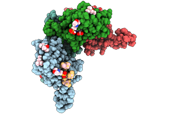 |
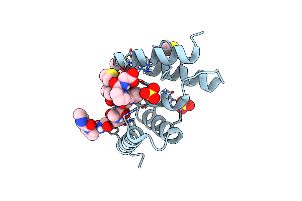
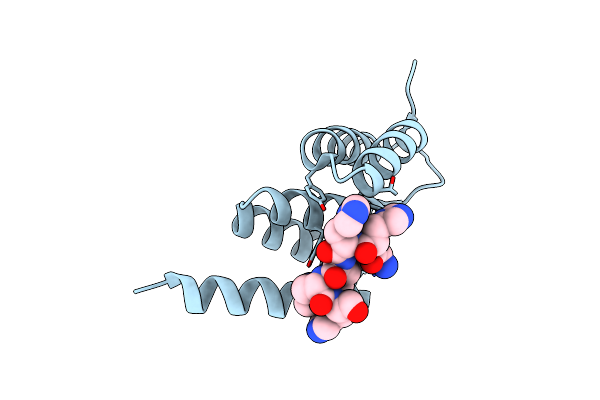
Peptide-Substrate-Binding (Psb) Domain Of Human Type I Collagen Prolyl 4-Hydroxylase Complexed With Pro-Pro-Gly-Pro-Arg-Gly-Pro-Pro-Gly.
Organism: Homo sapiens
Method: X-RAY DIFFRACTION Release Date: 2025-09-24 Classification: PROTEIN BINDING Ligands: MG, MPD, MPO, GLY |
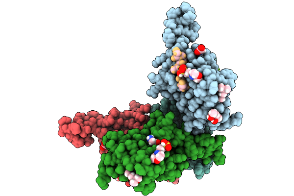 |
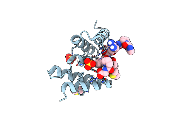
Peptide-Substrate-Binding (Psb) Domain Of Human Type I Collagen Prolyl 4-Hydroxylase Complexed With Pro-Hyp-Gly-Pro-Ala-Gly-Pro-Hyp-Gly.
Organism: Homo sapiens
Method: X-RAY DIFFRACTION Release Date: 2025-09-24 Classification: PROTEIN BINDING Ligands: GLY, MG, MPO, MPD |
 |
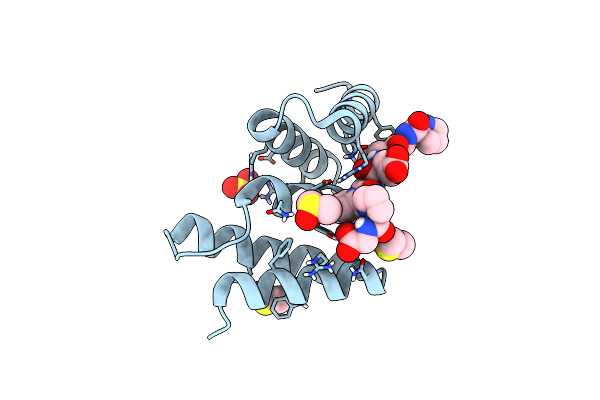
Peptide-Substrate-Binding (Psb) Domain Of Human Type I Collagen Prolyl 4-Hydroxylase Complexed With Pro-Pro-Gly-Pro-Ala-Gly-Pro-Pro-Gly.
Organism: Homo sapiens
Method: X-RAY DIFFRACTION Release Date: 2025-09-24 Classification: PROTEIN BINDING Ligands: GLY, MG, MPO, MPD |
 |
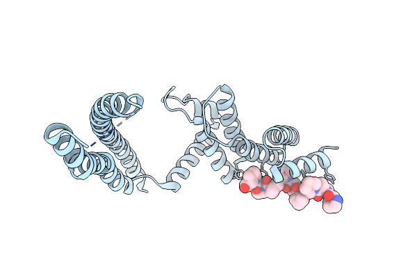
Peptide-Substrate-Binding (Psb) Domain Of Human Type Ii Collagen Prolyl 4-Hydroxylase Complexed With Pro-Hyp-Gly-Pro-Ala-Gly-Pro-Hyp-Gly.
Organism: Homo sapiens
Method: X-RAY DIFFRACTION Release Date: 2025-09-24 Classification: PROTEIN BINDING Ligands: GLY, SO4 |
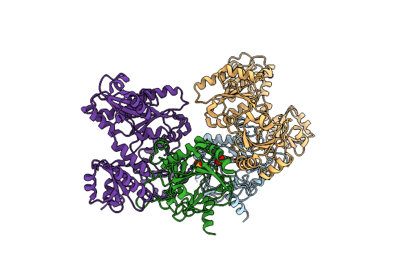 |
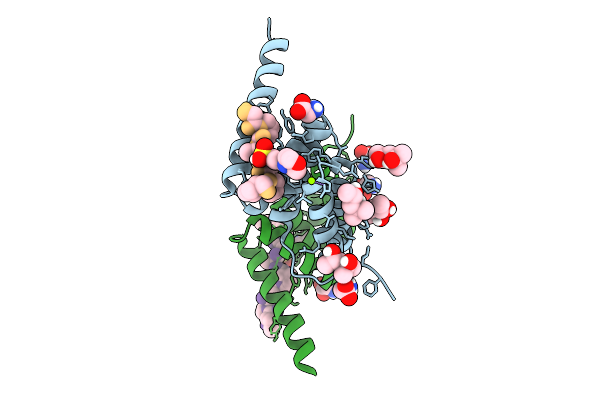
Crystal Structure Of The Heterodimeric Human C-P4H-Ii With Truncated Alpha Subunit (C-P4H-Ii Delta281)
Organism: Homo sapiens
Method: X-RAY DIFFRACTION Resolution:3.85 Å Release Date: 2022-11-09 Classification: HYDROLASE Ligands: SO4 |
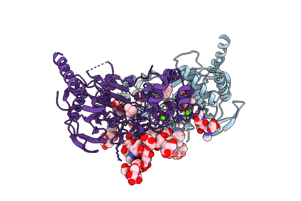 |
Organism: Homo sapiens
Method: X-RAY DIFFRACTION Resolution:2.25 Å Release Date: 2020-12-23 Classification: OXIDOREDUCTASE Ligands: OGA, FE2, TBU, GLY, CA, CL |
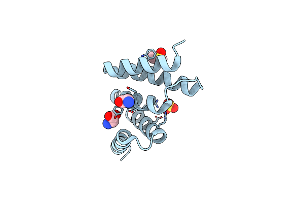 |
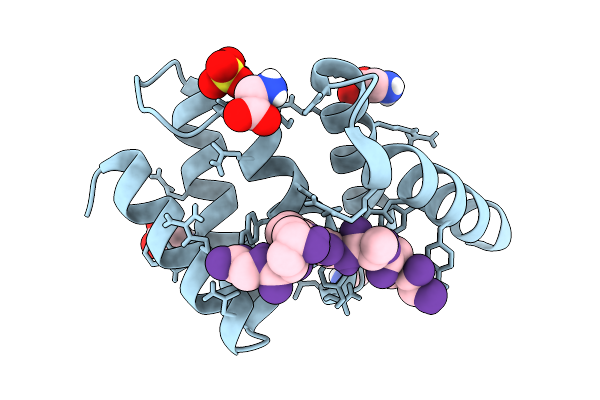
Crystal Structure Of An Unlignaded Peptide-Substrate-Binding Domain Of Human Type Ii Collagen Prolyl 4-Hydroxylase
Organism: Homo sapiens
Method: X-RAY DIFFRACTION Resolution:1.87 Å Release Date: 2018-09-12 Classification: HYDROLASE Ligands: SO4, DMS, GLY |
 |
Crystal Structure Of A Pro-9 Complexed Peptide-Substrate-Binding Domain Of Human Type Ii Collagen Prolyl 4-Hydroxylase
Organism: Homo sapiens, Synthetic construct
Method: X-RAY DIFFRACTION Resolution:2.00 Å Release Date: 2018-09-12 Classification: HYDROLASE Ligands: SO4, DMS |
 |
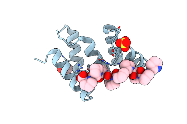
Crystal Structure Of Peptide-Substrate-Binding Domain Of Human Type Ii Collagen Prolyl 4-Hydroxylase Complex With Pro-Pro-Gly-Pro-Ala-Gly-Pro-Pro-Gly.
Organism: Homo sapiens, Synthetic construct
Method: X-RAY DIFFRACTION Resolution:1.48 Å Release Date: 2018-09-12 Classification: HYDROLASE Ligands: SO4, DMS |
 |
Crystal Structure The Peptide-Substrate-Binding Domain Of Human Type Ii Collagen Prolyl 4-Hydroxylase Complexed With Pro-Pro-Gly-Pro-Arg-Gly-Pro-Pro-Gly.
Organism: Homo sapiens, Synthetic construct
Method: X-RAY DIFFRACTION Resolution:1.55 Å Release Date: 2018-09-12 Classification: HYDROLASE Ligands: SO4, DMS |
 |
Crystal Structure The Peptide-Substrate-Binding Domain Of Human Type Ii Collagen Prolyl 4-Hydroxylase Complexed With Pro-Pro-Gly-Pro-Glu-Gly-Pro-Pro-Gly.
Organism: Homo sapiens, Synthetic construct
Method: X-RAY DIFFRACTION Resolution:1.68 Å Release Date: 2018-09-12 Classification: HYDROLASE Ligands: SO4, DMS |
 |
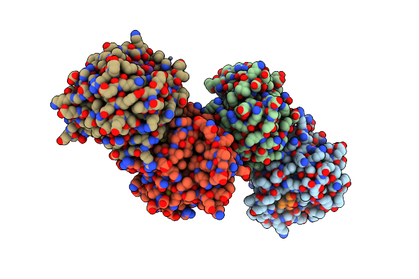
Crystal Structure Of Human Fibrillar Procollagen Type I C-Propeptide Homo-Trimer
Organism: Homo sapiens
Method: X-RAY DIFFRACTION Resolution:2.20 Å Release Date: 2017-03-22 Classification: STRUCTURAL PROTEIN Ligands: CA, GOL, CL |
 |
Crystal Structure Of The Semet-Labeled N-Terminal Domain And Peptide Substrate Binding Domain Of Alpha Subunit Of Prolyl-4 Hydroxylase Type I From Human.
Organism: Homo sapiens
Method: X-RAY DIFFRACTION Resolution:2.99 Å Release Date: 2013-10-09 Classification: OXIDOREDUCTASE |
 |
Crystal Structure Of The Apo Form Of N-Terminal Domain And Peptide Substrate Binding Domain Of Prolyl-4 Hydroxylase Type I From Human
Organism: Homo sapiens
Method: X-RAY DIFFRACTION Resolution:2.20 Å Release Date: 2013-10-09 Classification: OXIDOREDUCTASE |
 |
Crystal Structure Of The Peptide(Pro-Pro-Gly)3 Bound Complex Of N- Terminal Domain And Peptide Substrate Binding Domain Of Prolyl-4 Hydroxylase (Residues 1-238) Type I From Human
Organism: Homo sapiens, Synthetic construct
Method: X-RAY DIFFRACTION Resolution:1.90 Å Release Date: 2013-10-09 Classification: OXIDOREDUCTASE |
 |
Crystal Structure Of The Peptide(Pro-Pro-Gly)3 Bound Complex Of N- Terminal Domain And Peptide Substrate Binding Domain Of Prolyl-4 Hydroxylase (Residues 1-244) Type I From Human
Organism: Homo sapiens
Method: X-RAY DIFFRACTION Resolution:2.95 Å Release Date: 2013-10-09 Classification: OXIDOREDUCTASE |
 |
Crystal Structure Of The Peptide(Pro)9 Bound Complex Of N-Terminal Domain And Peptide Substrate Binding Domain Of Prolyl-4 Hydroxylase (Residues 1-238) Type I From Human
Organism: Homo sapiens, Synthetic construct
Method: X-RAY DIFFRACTION Resolution:1.90 Å Release Date: 2013-10-09 Classification: OXIDOREDUCTASE |
 |
Crystal Structure Of Wild Type Peptide-Binding Domain Of Human Type I Collagen Prolyl 4-Hydroxylase.
Organism: Homo sapiens, Synthetic construct
Method: X-RAY DIFFRACTION Resolution:2.03 Å Release Date: 2009-11-17 Classification: OXIDOREDUCTASE |
 |
Algal Prolyl 4-Hydroxylase Complexed With Zinc And (Ser-Pro)5 Peptide Substrate
Organism: Chlamydomonas reinhardtii
Method: X-RAY DIFFRACTION Resolution:1.98 Å Release Date: 2009-06-23 Classification: HYDROLASE Ligands: ZN, ACY |
 |
Crystal Structure Of Chlamydomonas Reinhardtii Prolyl-4 Hydroxylase Type I Complexed With Zinc And Pyridine-2,4-Dicarboxylate
Organism: Chlamydomonas reinhardtii
Method: X-RAY DIFFRACTION Resolution:1.85 Å Release Date: 2007-10-30 Classification: HYDROLASE Ligands: ZN, PD2, GOL, SO4 |