
Search Count: 11
 |
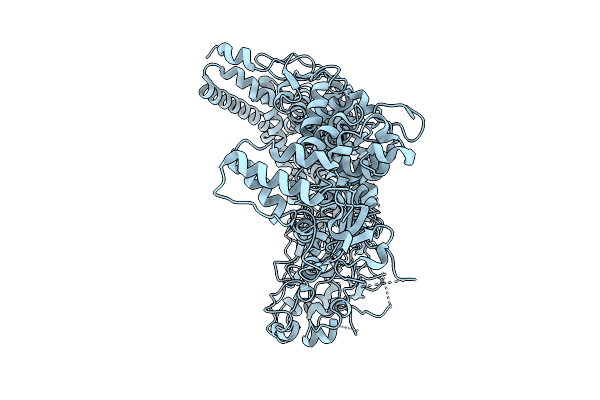
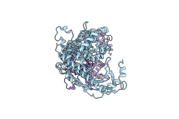
Cryo-Em Structure Of Essential Mycoplasma Pneumoniae Lipoprotein Mpn444 Homotrimer
Organism: Mycoplasmoides pneumoniae m129
Method: ELECTRON MICROSCOPY Release Date: 2025-09-17 Classification: UNKNOWN FUNCTION |
 |
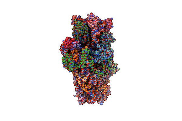
Organism: Mycoplasmoides pneumoniae m129
Method: ELECTRON MICROSCOPY Release Date: 2025-09-10 Classification: UNKNOWN FUNCTION |
 |
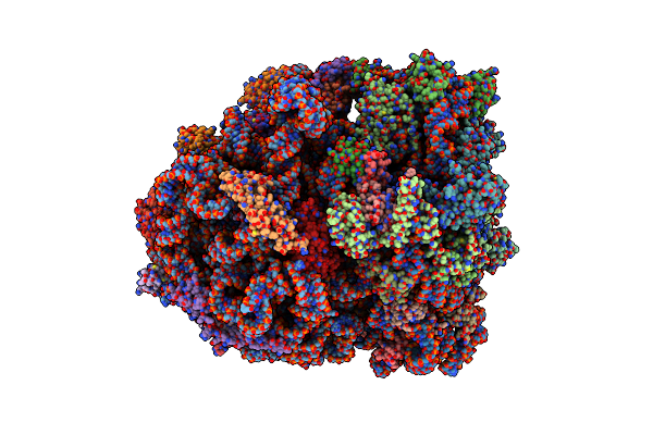
Single-Particle Cryo-Em Of Mycoplasma Pneumoniae Adhesin P1 Complexed With The Anti-Adhesive Fab Fragment.
Organism: Mycoplasmoides pneumoniae m129, Mus musculus
Method: ELECTRON MICROSCOPY Release Date: 2025-03-05 Classification: CELL ADHESION |
 |
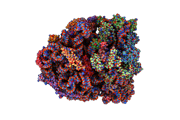
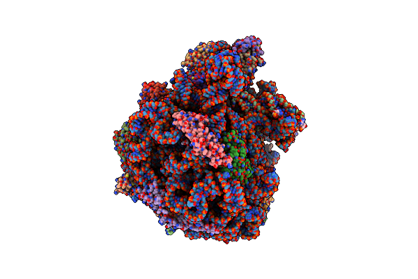
Mycoplasma Pneumoniae Small Ribosomal Subunit In Chloramphenicol-Treated Cells
Organism: Mycoplasmoides pneumoniae m129
Method: ELECTRON MICROSCOPY Release Date: 2024-11-20 Classification: TRANSLATION Ligands: SPD, PUT, N2P, MG, ZN |
 |
Organism: Mycoplasmoides pneumoniae m129
Method: ELECTRON MICROSCOPY Release Date: 2024-11-20 Classification: TRANSLATION Ligands: ZN, CLM, K, MG, PUT, SPM, SPD, N2P, LYS |
 |
Organism: Mycoplasmoides pneumoniae m129
Method: ELECTRON MICROSCOPY Release Date: 2024-11-20 Classification: TRANSLATION Ligands: ZN, CLM, K, MG, PUT, SPM, SPD, N2P, LYS |
 |
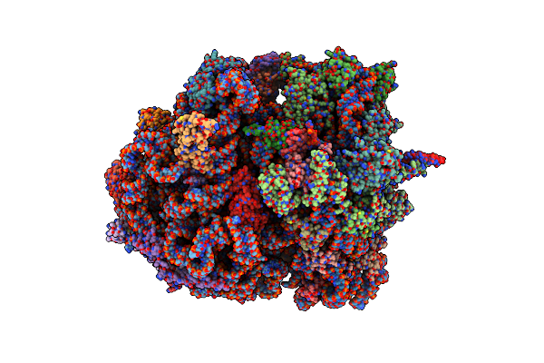
Mycoplasma Pneumoniae Large Ribosomal Subunit In Chloramphenicol-Treated Cells
Organism: Mycoplasmoides pneumoniae m129
Method: ELECTRON MICROSCOPY Release Date: 2024-11-20 Classification: TRANSLATION Ligands: ZN, CLM, K, MG, PUT, SPM, SPD, N2P, LYS |
 |
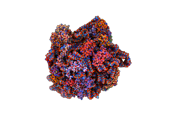
Mycoplasma Pneumoniae Di-Ribosome In Chloramphenicol-Treated Cells (Leading 70S)
Organism: Mycoplasmoides pneumoniae m129
Method: ELECTRON MICROSCOPY Release Date: 2024-11-20 Classification: TRANSLATION Ligands: ZN, CLM, K, MG, LYS |
 |
Mycoplasma Pneumoniae Di-Ribosome In Chloramphenicol-Treated Cells (Following 70S)
Organism: Mycoplasmoides pneumoniae m129
Method: ELECTRON MICROSCOPY Release Date: 2024-11-20 Classification: TRANSLATION Ligands: ZN, CLM, K, MG |
 |
P116 Dimer In The Full State (Pdb Structure Of The Full-Length Ectodomain Truncated To Amino Acids 246-818)
Organism: Mycoplasmoides pneumoniae m129
Method: ELECTRON MICROSCOPY Release Date: 2024-05-29 Classification: LIPID BINDING PROTEIN |
 |
Organism: Mycoplasmoides pneumoniae m129, Mycoplasma pneumoniae m129
Method: ELECTRON MICROSCOPY Release Date: 2022-05-25 Classification: TRANSLATION |