
Search Count: 30
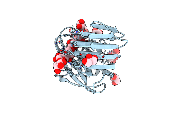 |
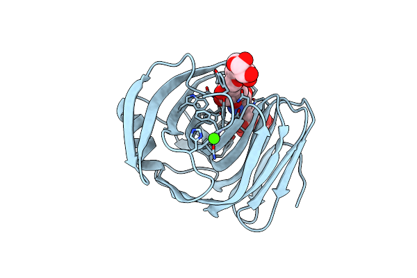
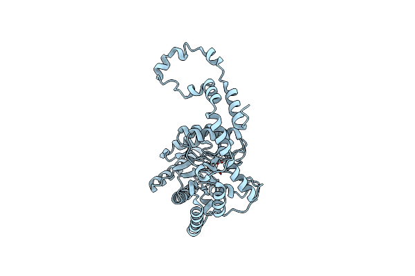
Crystal Structures Of Mutant Endo- -1,4-Xylanase Ii Complexed With Substrate (1.15 A) And Products (1.6 A)
Organism: Trichoderma reesei
Method: X-RAY DIFFRACTION Resolution:1.15 Å Release Date: 2014-01-08 Classification: HYDROLASE Ligands: GOL, CIT |
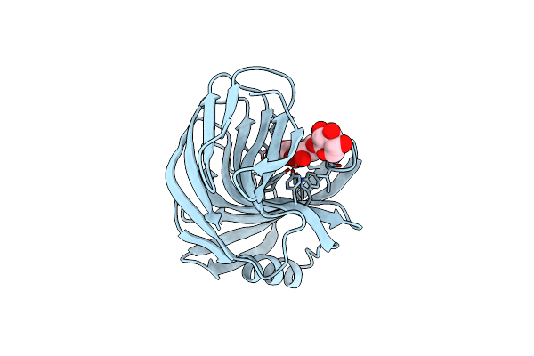 |
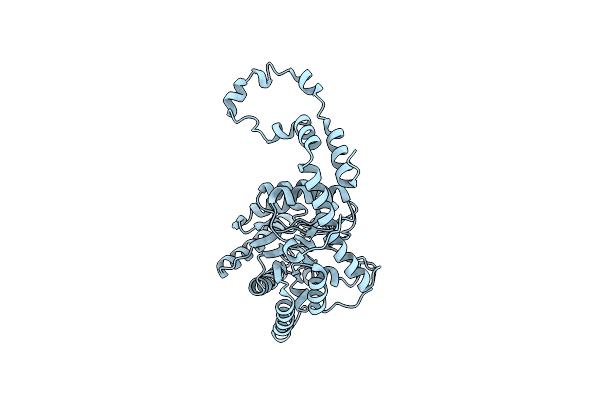
Crystal Structures Of Mutant Endo-Beta-1,4-Xylanase Ii Complexed With Substrate (1.15 A) And Products (1.6 A)
Organism: Trichoderma reesei
Method: X-RAY DIFFRACTION Resolution:1.55 Å Release Date: 2014-01-08 Classification: HYDROLASE |
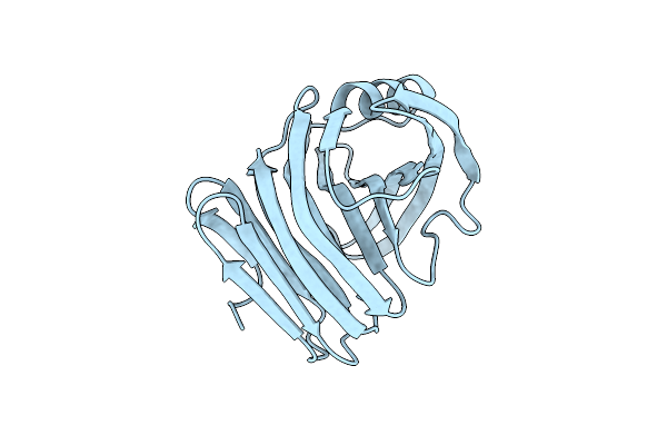 |
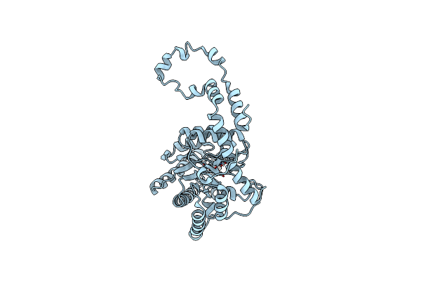
Crystal Structures Of Mutant Endo-Beta-1,4-Xylanase Ii Complexed With Substrate (1.15 A) And Products (1.6 A)
Organism: Trichoderma reesei
Method: X-RAY DIFFRACTION Resolution:1.10 Å Release Date: 2014-01-08 Classification: HYDROLASE Ligands: IOD |
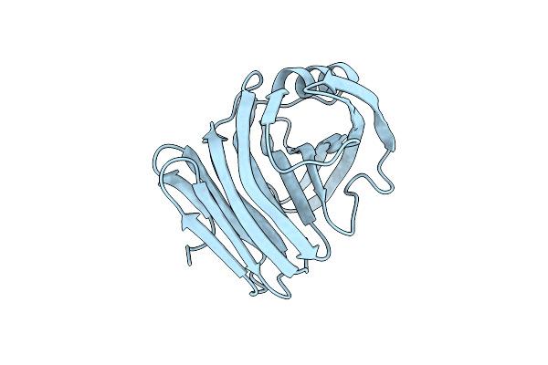 |
Crystal Structures Of Mutant Endo-Beta-1,4-Xylanase Ii (E177Q) In The Apo Form
Organism: Trichoderma reesei
Method: X-RAY DIFFRACTION Resolution:1.50 Å Release Date: 2014-01-08 Classification: HYDROLASE Ligands: IOD |
 |
Crystal Structures Of Mutant Endo-Beta-1,4-Xylanase Ii Complexed With Substrate And Products
Organism: Trichoderma reesei
Method: X-RAY DIFFRACTION Resolution:1.65 Å Release Date: 2014-01-08 Classification: HYDROLASE Ligands: TRS, CA |
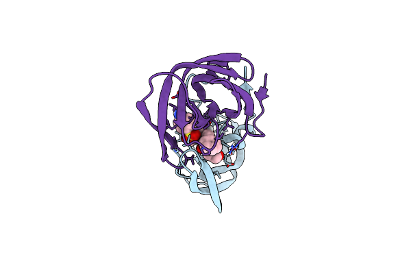 |
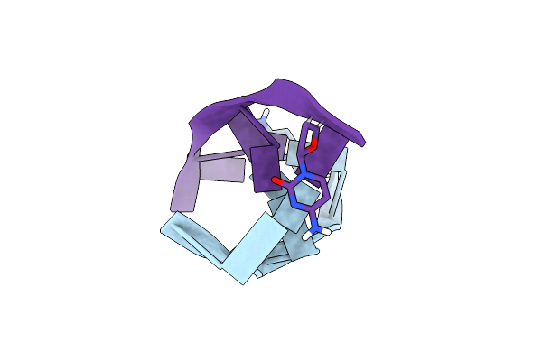
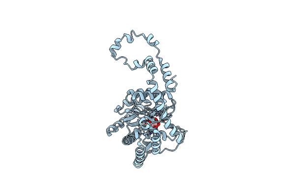
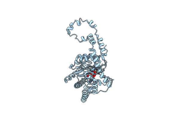
Joint Neutron And X-Ray Structure Of Per-Deuterated Hiv-1 Protease In Complex With Clinical Inhibitor Amprenavir
Organism: Human immunodeficiency virus type 1
Method: NEUTRON DIFFRACTION, X-RAY DIFFRACTION Resolution:2.01 Å, 2.00 Å Release Date: 2013-07-24 Classification: HYDROLASE/HYDROLASE INHIBITOR Ligands: CL, 478, DOD |
 |
Room-Temperature X-Ray Structure Of D-Xylose Isomerase In Complex With 2Mg2+ Ions And Xylitol At Ph 7.7
Organism: Streptomyces rubiginosus
Method: X-RAY DIFFRACTION Resolution:2.00 Å Release Date: 2012-08-29 Classification: isomerase/isomerase inhibitor Ligands: MG, XYL |
 |
Room-Temperature Joint X-Ray/Neutron Structure Of D-Xylose Isomerase In Complex With 2Ni2+ And Per-Deuterated D-Sorbitol At Ph 5.9
Organism: Streptomyces rubiginosus
Method: NEUTRON DIFFRACTION, X-RAY DIFFRACTION Resolution:2.00 Å Release Date: 2012-08-29 Classification: ISOMERASE Ligands: NI, SOR, DOD |
 |
Room Temperature Ultra-High Resolution Time-Of-Flight Neutron And X-Ray Diffraction Studies Of H/D Exchanged Crambin
Organism: Crambe hispanica subsp. abyssinica
Method: X-RAY DIFFRACTION Resolution:0.85 Å Release Date: 2012-02-08 Classification: PLANT PROTEIN |
 |
Organism: Homo sapiens
Method: NEUTRON DIFFRACTION, X-RAY DIFFRACTION Resolution:2.00 Å Release Date: 2011-11-09 Classification: LYASE Ligands: ZN, DOD |
 |
Room Temperature X-Ray Structure Of D-Xylose Isomerase In Complex With 0.6Ni2+ Cation Bound In M2 Metal Binding Site At Ph=5.8
Organism: Streptomyces rubiginosus
Method: X-RAY DIFFRACTION Resolution:1.85 Å Release Date: 2011-08-17 Classification: ISOMERASE Ligands: NI |
 |
Organism: Streptomyces rubiginosus
Method: NEUTRON DIFFRACTION, X-RAY DIFFRACTION Resolution:2.0 Å, 1.7 Å Release Date: 2011-08-17 Classification: ISOMERASE Ligands: D8U, DOD |
 |
Reintroducing Electrostatics Into Macromolecular Crystallographic Refinement: Z-Dna (X-Ray)
Method: X-RAY DIFFRACTION, NEUTRON DIFFRACTION
Resolution:1.53 Å, 1.4 Å Release Date: 2011-03-02 Classification: DNA Ligands: DOD |
 |
Organism: Streptomyces rubiginosus
Method: X-RAY DIFFRACTION Resolution:2.00 Å Release Date: 2010-09-29 Classification: ISOMERASE |
 |
Room Temperature Neutron Structure Of Apo-D-Xylose Isomerase (Refined Jointly With X-Ray Structure 3Kbj)
Organism: Streptomyces rubiginosus
Method: NEUTRON DIFFRACTION, X-RAY DIFFRACTION Resolution:1.8 Å, 2.0 Å Release Date: 2010-09-29 Classification: ISOMERASE Ligands: DOD |
 |
Room Temperature X-Ray Structure Of D-Xylose Isomerase Complexed With 2Cd(2+) Co-Factors And D12-D-Alpha-Glucose In The Cyclic Form
Organism: Streptomyces rubiginosus
Method: X-RAY DIFFRACTION Resolution:2.00 Å Release Date: 2010-06-16 Classification: ISOMERASE Ligands: CD, GLC |
 |
Room Temperature Structure Of D-Xylose Isomerase In Complex With 2Ni(2+) Co-Factors And D12-D-Glucose In The Linear Form
Organism: Streptomyces rubiginosus
Method: X-RAY DIFFRACTION Resolution:1.53 Å Release Date: 2010-06-16 Classification: ISOMERASE Ligands: NI, GLO |
 |
Room Temperature X-Ray Structure Of D-Xylose Isomerase In Complex With 2Cd(2+) Co-Factors
Organism: Streptomyces rubiginosus
Method: X-RAY DIFFRACTION Resolution:1.80 Å Release Date: 2010-06-16 Classification: ISOMERASE Ligands: CD |
 |
Room Temperature Structure Of D-Xylose Isomerase In Complex With 2Ni(2+) Co-Factors
Organism: Streptomyces rubiginosus
Method: X-RAY DIFFRACTION Resolution:1.80 Å Release Date: 2010-06-16 Classification: ISOMERASE Ligands: NI |
 |
Room Temperature X-Ray Mixed-Metal Structure Of D-Xylose Isomerase In Complex With Ni(2+) And Mg(2+) Co-Factors
Organism: Streptomyces rubiginosus
Method: X-RAY DIFFRACTION Resolution:1.60 Å Release Date: 2010-06-16 Classification: ISOMERASE Ligands: NI, MG |