
Search Count: 67
 |
Organism: Homo sapiens
Method: ELECTRON MICROSCOPY Resolution:3.10 Å Release Date: 2025-12-10 Classification: LIGASE Ligands: ZN |
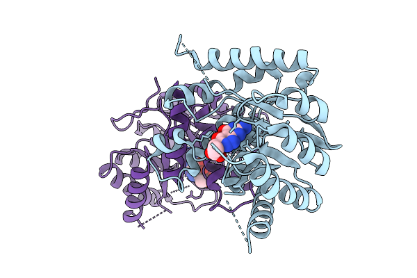 |
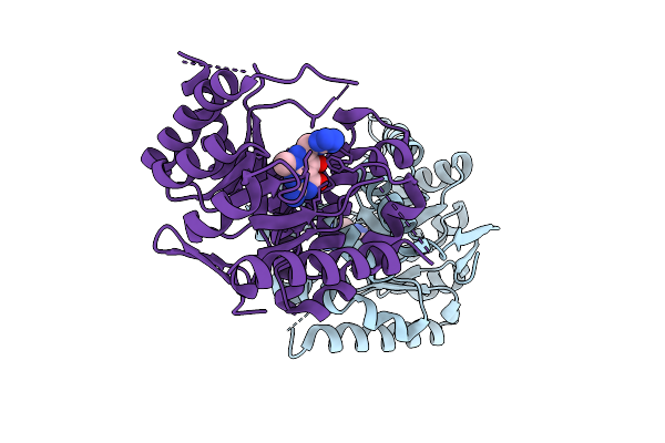
Organism: Escherichia coli
Method: X-RAY DIFFRACTION Resolution:3.00 Å Release Date: 2025-11-05 Classification: TRANSFERASE Ligands: A1CG3 |
 |
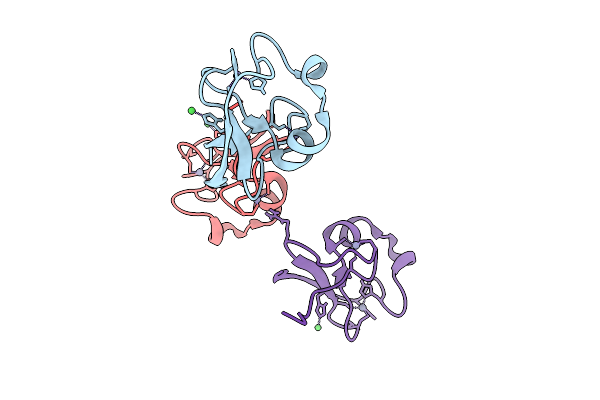
Organism: Escherichia coli
Method: X-RAY DIFFRACTION Resolution:2.14 Å Release Date: 2025-11-05 Classification: TRANSFERASE Ligands: A1CG7 |
 |
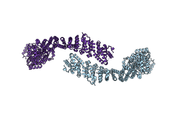
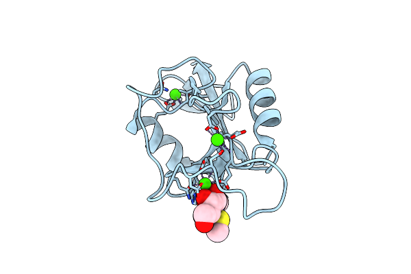
R-Degron Fused Zz-Domain Of The Arabidopsis Thaliana E3 Ubiquitin-Protein Ligase Big
Organism: Arabidopsis thaliana
Method: X-RAY DIFFRACTION Resolution:1.50 Å Release Date: 2025-09-17 Classification: LIGASE Ligands: ZN, NI |
 |
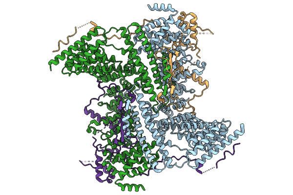
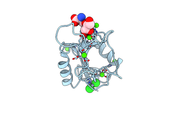
Cryo-Em Structure Of The Core Of The Arabidopsis Thaliana Ubr4/Di19/Calm1 Complex
Organism: Arabidopsis thaliana
Method: ELECTRON MICROSCOPY Resolution:3.90 Å Release Date: 2025-09-03 Classification: LIGASE |
 |
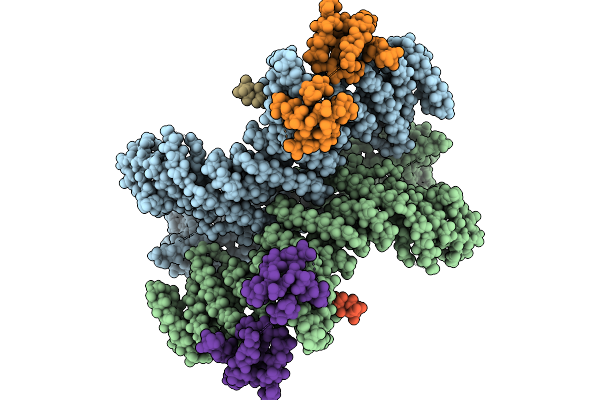
Cryo-Em Structure Of The Human Ubr4/Kcmf1/Calm1 Complex (C-Term Dimer Interface Focused Refinement)
Organism: Homo sapiens
Method: ELECTRON MICROSCOPY Resolution:3.10 Å Release Date: 2025-09-03 Classification: LIGASE Ligands: ZN |
 |
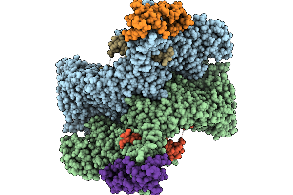
Cryo-Em Structure Of The Human Ubr4/Kcmf1/Calm1 Complex (Calm1 Focused Refinement)
Organism: Homo sapiens
Method: ELECTRON MICROSCOPY Resolution:3.10 Å Release Date: 2025-09-03 Classification: LIGASE Ligands: ZN |
 |
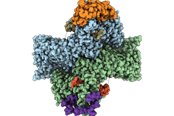
Cryo-Em Structure Of The Human Ubr4/Kcmf1/Calm1 Complex (N-Term Focused Refinement)
Organism: Homo sapiens
Method: ELECTRON MICROSCOPY Resolution:3.60 Å Release Date: 2025-09-03 Classification: LIGASE |
 |
Cryo-Em Structure Of The Human Ubr4/Kcmf1/Calm1 Complex (Bp Focused Refinement)
Organism: Homo sapiens
Method: ELECTRON MICROSCOPY Resolution:4.80 Å Release Date: 2025-09-03 Classification: LIGASE |
 |
Cryo-Em Structure Of The Human Ubr4/Kcmf1/Calm1 Complex (C-Term Focused Refinement)
Organism: Homo sapiens
Method: ELECTRON MICROSCOPY Resolution:3.40 Å Release Date: 2025-09-03 Classification: LIGASE |
 |
Cryo-Em Structure Of The C. Elegans Ubr4/Kcmf1 Complex (C-Term Dimer Interface Focused Refinement)
Organism: Caenorhabditis elegans
Method: ELECTRON MICROSCOPY Resolution:2.60 Å Release Date: 2025-09-03 Classification: LIGASE Ligands: ZN |
 |
Cryo-Em Structure Of The C. Elegans Ubr4/Kcmf1 Complex (N-Term Focused Refinement)
Organism: Caenorhabditis elegans
Method: ELECTRON MICROSCOPY Resolution:5.70 Å Release Date: 2025-09-03 Classification: LIGASE |
 |
Cryo-Em Structure Of The C. Elegans Ubr4/Kcmf1 Complex (Side Focused Refinement)
Organism: Caenorhabditis elegans
Method: ELECTRON MICROSCOPY Resolution:3.50 Å Release Date: 2025-09-03 Classification: LIGASE |
 |
Organism: Homo sapiens
Method: X-RAY DIFFRACTION Resolution:1.92 Å Release Date: 2025-08-27 Classification: PEPTIDE BINDING PROTEIN Ligands: ZN |
 |
Organism: Homo sapiens, Synthetic construct
Method: X-RAY DIFFRACTION Resolution:1.71 Å Release Date: 2025-08-27 Classification: LIGASE Ligands: ZN |
 |
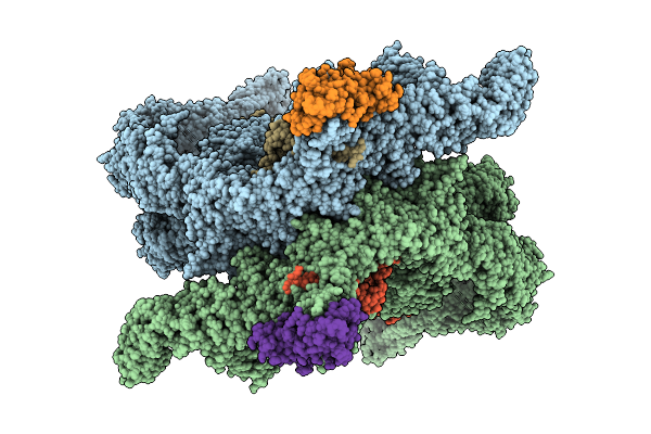
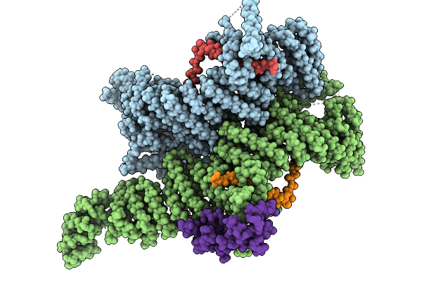
Crystal Structure Of The Wild-Type Thermus Thermophilus 70S Ribosome In Complex With Spectinomycin, Mrna, Deacylated A- And E-Site Trnaphe, And Deacylated P-Site Trnamet At 2.60A Resolution
Organism: Escherichia coli, Escherichia phage t4, Thermus thermophilus hb8
Method: X-RAY DIFFRACTION Resolution:2.60 Å Release Date: 2024-08-07 Classification: RIBOSOME/INHIBITOR,ANTIBIOTIC Ligands: MG, K, ZN, SCM, SF4 |
 |
Crystal Structure Of The Wild-Type Thermus Thermophilus 70S Ribosome In Complex With Spectinomycin Derivative 2694, Mrna, Deacylated A- And E-Site Trnaphe, And Deacylated P-Site Trnamet At 2.75A Resolution
Organism: Escherichia coli, Escherichia phage t4, Thermus thermophilus hb8
Method: X-RAY DIFFRACTION Resolution:2.75 Å Release Date: 2024-08-07 Classification: RIBOSOME/INHIBITOR Ligands: MG, ZN, Y7K, SF4, K |
 |
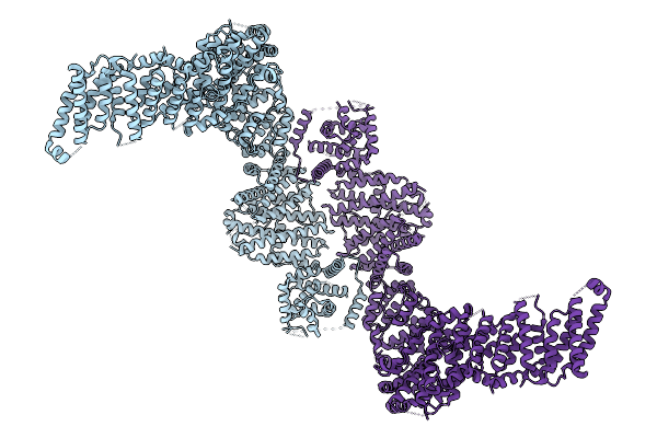
Organism: Nematocida sp. ertm5
Method: X-RAY DIFFRACTION Resolution:3.04 Å Release Date: 2021-07-28 Classification: LIGASE |
 |
Crystal Structure Of The Carbohydrate Recognition Domain Of The Human Macrophage Galactose C-Type Lectin Bound To Methyl 2-(Acetylamino)-2-Deoxy-1-Thio-Alpha-D-Galactopyranose
Organism: Homo sapiens
Method: X-RAY DIFFRACTION Resolution:2.31 Å Release Date: 2021-03-31 Classification: SIGNALING PROTEIN Ligands: Q3M, CA, CL |
 |
Crystal Structure Of The Carbohydrate Recognition Domain Of The Human Macrophage Galactose C-Type Lectin Bound To The Tumor-Associated Tn Antigen
Organism: Homo sapiens
Method: X-RAY DIFFRACTION Resolution:2.00 Å Release Date: 2021-03-10 Classification: SIGNALING PROTEIN Ligands: CA, CL, ZN, SER, A2G |