
Search Count: 166
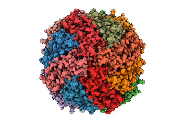 |
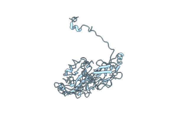
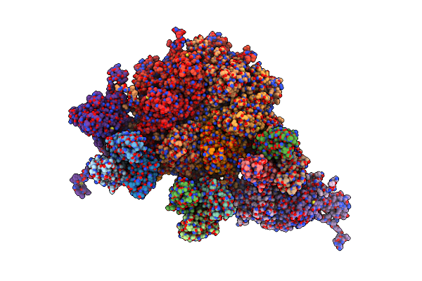
Apoferritin (118% Super Resolution Nyquist, 236% Physical Nyquist) By Pasr On Acquisition-Time Super Resolution K3 Data
Organism: Mus musculus
Method: ELECTRON MICROSCOPY Release Date: 2025-09-17 Classification: METAL BINDING PROTEIN Ligands: FE |
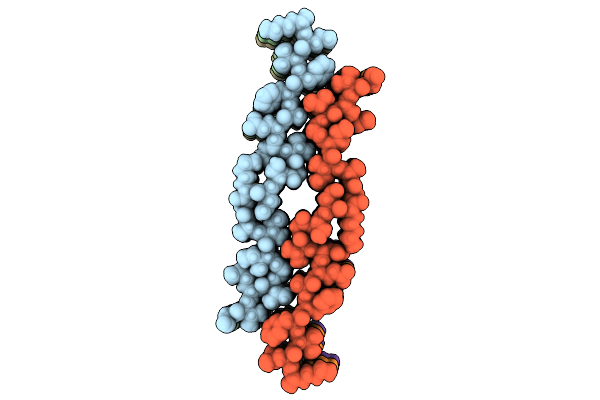 |
Organism: Homo sapiens
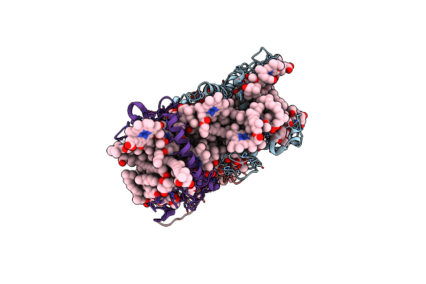
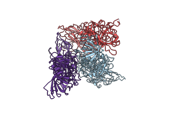
Method: ELECTRON MICROSCOPY Release Date: 2025-07-09 Classification: PROTEIN FIBRIL |
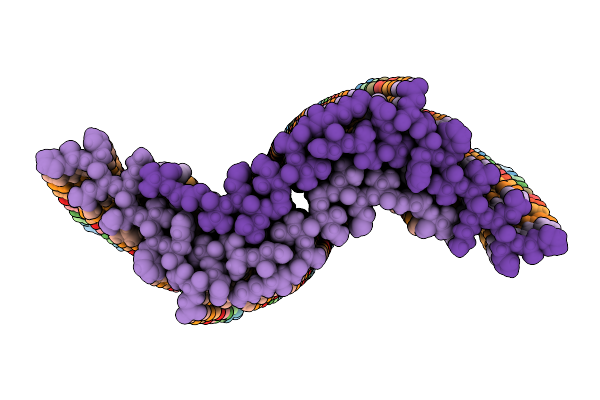 |
Organism: Homo sapiens
Method: ELECTRON MICROSCOPY Release Date: 2025-07-09 Classification: PROTEIN FIBRIL |
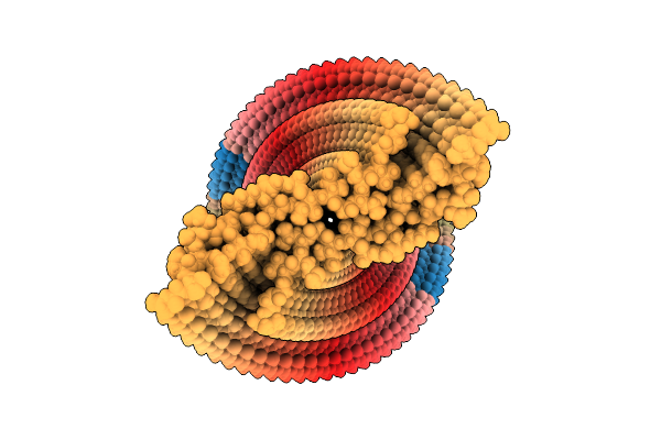 |
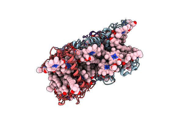
Organism: Homo sapiens
Method: ELECTRON MICROSCOPY Release Date: 2025-07-09 Classification: PROTEIN FIBRIL |
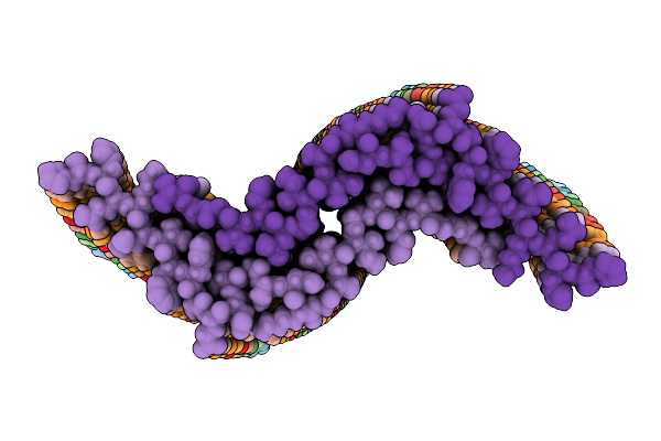 |
Organism: Homo sapiens
Method: ELECTRON MICROSCOPY Release Date: 2025-07-09 Classification: PROTEIN FIBRIL |
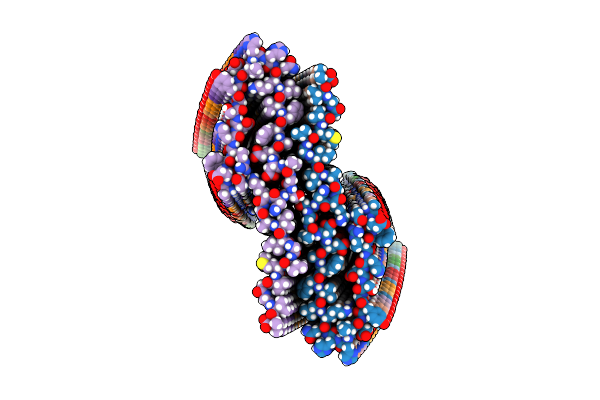 |
Organism: Homo sapiens
Method: ELECTRON MICROSCOPY Release Date: 2025-02-26 Classification: PROTEIN FIBRIL |
 |
Crystal Structure Of Horse-Spleen L-Ferritin Fused With Amyloid Beta Peptide (1-42).
Organism: Equus caballus
Method: X-RAY DIFFRACTION Resolution:2.00 Å Release Date: 2024-04-03 Classification: METAL BINDING PROTEIN Ligands: CD, SO4, CL |
 |
Organism: Spirochaeta thermophila dsm 6578, Corynebacterium pollutisoli
Method: X-RAY DIFFRACTION Resolution:3.34 Å Release Date: 2024-03-27 Classification: TRANSPORT PROTEIN Ligands: THJ, OLC |
 |
Organism: Spirochaeta thermophila dsm 6578, Corynebacterium pollutisoli
Method: X-RAY DIFFRACTION Resolution:2.60 Å Release Date: 2024-03-27 Classification: TRANSPORT PROTEIN Ligands: OLC |
 |
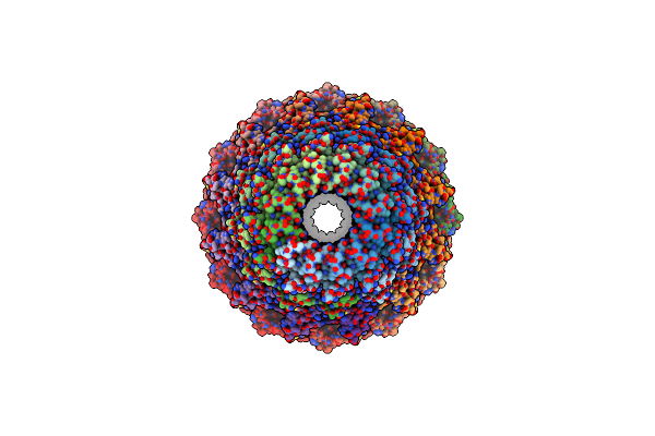
Organism: Staphylococcus phage s6
Method: ELECTRON MICROSCOPY Release Date: 2023-12-06 Classification: VIRUS |
 |
Organism: Melbournevirus
Method: ELECTRON MICROSCOPY Release Date: 2023-08-02 Classification: VIRAL PROTEIN |
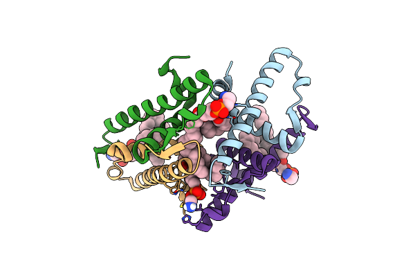 |
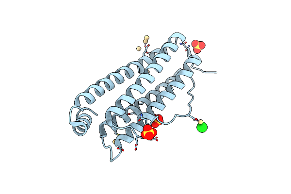
Organism: Schizosaccharomyces pombe (strain 972 / atcc 24843)
Method: X-RAY DIFFRACTION Resolution:1.58 Å Release Date: 2023-05-17 Classification: LIPID BINDING PROTEIN Ligands: 3PE |
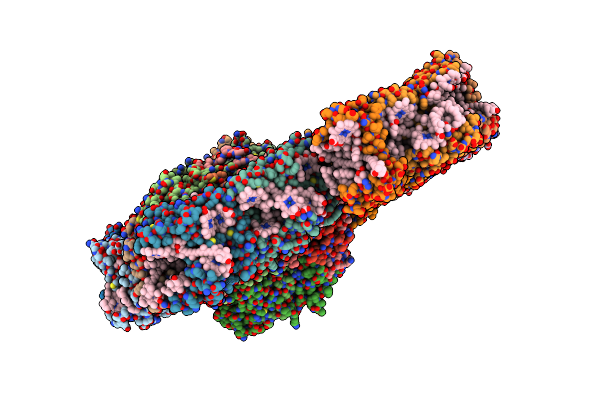 |
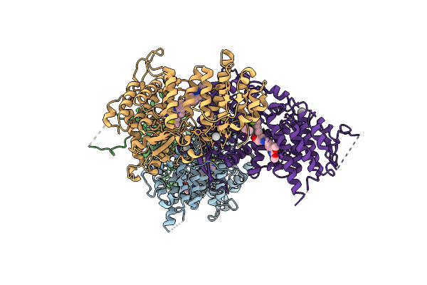
Cryo-Em Structure Of The Psi-Lhci-Lhcp Supercomplex From Ostreococcus Tauri
Organism: Ostreococcus tauri
Method: ELECTRON MICROSCOPY Release Date: 2023-04-26 Classification: PHOTOSYNTHESIS Ligands: CHL, CLA, IWJ, XAT, LHG, Q6L, BCR, LMG, SQD, CL0, PQN, SF4, DGD, NEX, KC2 |
 |
Organism: Ostreococcus tauri
Method: ELECTRON MICROSCOPY Release Date: 2023-04-26 Classification: ELECTRON TRANSPORT Ligands: CLA, CHL, KC2, Q6L, NEX, IWJ |
 |
Cryo-Em Structure Of The Prasinophyte-Specific Light-Harvesting Complex (Lhcp)From Ostreococcus Tauri
Organism: Ostreococcus tauri
Method: ELECTRON MICROSCOPY Release Date: 2023-04-26 Classification: ELECTRON TRANSPORT Ligands: CLA, CHL, KC2, Q6L, NEX, IWJ |
 |
Cryo-Em Structure Of The Prasinophyte-Specific Light-Harvesting Complex (Lhcp)From Ostreococcus Tauri
Organism: Ostreococcus tauri
Method: ELECTRON MICROSCOPY Release Date: 2023-04-26 Classification: ELECTRON TRANSPORT Ligands: CLA, CHL, KC2, Q6L, IWJ, NEX |
 |
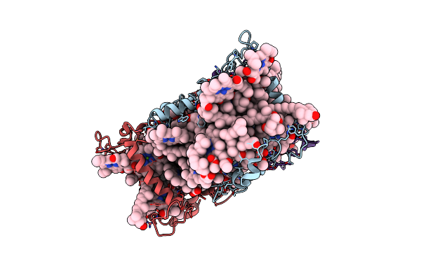
Organism: Homo sapiens
Method: X-RAY DIFFRACTION Resolution:2.60 Å Release Date: 2023-03-22 Classification: TRANSCRIPTION/INHIBITOR Ligands: 7IX, UNX, DMS |
 |
Organism: Helicobacter phage khp30
Method: ELECTRON MICROSCOPY Release Date: 2023-03-01 Classification: VIRAL PROTEIN |
 |
Organism: Sapovirus hu/gi.6/nichinan/fp05284/2005/jpn
Method: ELECTRON MICROSCOPY Release Date: 2021-12-15 Classification: VIRUS |
 |
Organism: Helicobacter pylori bacteriophage khp30
Method: ELECTRON MICROSCOPY Release Date: 2021-10-27 Classification: VIRUS |