
Search Count: 10
 |
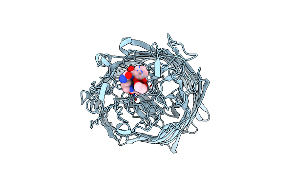
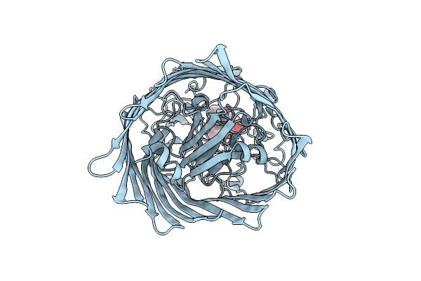
Crystal Structure Of The Ferric Enterobactin Receptor (Pfea) From Pseudomonas Aeruginosa In Complex With Tcv-L6
Organism: Pseudomonas aeruginosa pao1
Method: X-RAY DIFFRACTION Resolution:2.66 Å Release Date: 2021-08-04 Classification: MEMBRANE PROTEIN Ligands: V82, FE |
 |
Crystal Structure Of The Ferric Enterobactin Receptor (Pfea) In Complex With Bcv-L5
Organism: Pseudomonas aeruginosa pao1
Method: X-RAY DIFFRACTION Resolution:3.04 Å Release Date: 2021-01-20 Classification: MEMBRANE PROTEIN Ligands: OWT, EDO, FE |
 |
Crystal Structure Of The Ferric Enterobactin Receptor (Pfea) In Complex With Bcv-L6
Organism: Pseudomonas aeruginosa (strain atcc 15692 / dsm 22644 / cip 104116 / jcm 14847 / lmg 12228 / 1c / prs 101 / pao1)
Method: X-RAY DIFFRACTION Resolution:3.03 Å Release Date: 2021-01-20 Classification: MEMBRANE PROTEIN Ligands: EDO, Q5N, FE |
 |
Crystal Structure Of The Ferric Enterobactin Receptor (Pfea) In Complex With Tcv_L5
Organism: Pseudomonas aeruginosa (strain atcc 15692 / dsm 22644 / cip 104116 / jcm 14847 / lmg 12228 / 1c / prs 101 / pao1)
Method: X-RAY DIFFRACTION Resolution:2.72 Å Release Date: 2020-07-29 Classification: MEMBRANE PROTEIN Ligands: FE, QDQ |
 |
Crystal Structure Of The Ferric Enterobactin Receptor (Pfea) In Complex With Bcv
Organism: Pseudomonas aeruginosa pao1
Method: X-RAY DIFFRACTION Resolution:2.71 Å Release Date: 2020-07-29 Classification: MEMBRANE PROTEIN Ligands: Q62, EDO, FE |
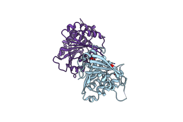 |
Crystal Structure Of The Ferric Enterobactin Esterase (Pfee) From Pseudomonas Aeruginosa
Organism: Pseudomonas aeruginosa
Method: X-RAY DIFFRACTION Resolution:2.00 Å Release Date: 2018-06-20 Classification: HYDROLASE Ligands: NO3 |
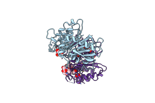 |
Crystal Structure Of The Ferric Enterobactin Esterase (Pfee) Mutant(S157A) From Pseudomonas Aeruginosa In Presence Of Enterobactin
Organism: Pseudomonas aeruginosa pao1
Method: X-RAY DIFFRACTION Resolution:1.66 Å Release Date: 2018-06-20 Classification: HYDROLASE Ligands: FE, DBS, NO3, EDO, EB4 |
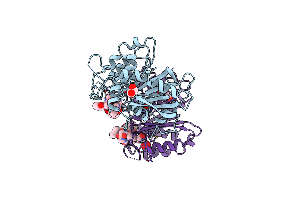 |
Crystal Structure Of The Ferric Enterobactin Esterase (Pfee) Mutant(S157A) From Pseudomonas Aeruginosa In Complex With Tris-Catechol Vector
Organism: Pseudomonas aeruginosa
Method: X-RAY DIFFRACTION Resolution:1.49 Å Release Date: 2018-06-20 Classification: HYDROLASE Ligands: FE, 8SW, NO3, EDO |
 |
Crystal Structure Of The Ferric Enterobactin Esterase (Pfee) From Pseudomonas Aeruginosa In Complex With The Tris-Catechol Vector
Organism: Pseudomonas aeruginosa
Method: X-RAY DIFFRACTION Resolution:3.11 Å Release Date: 2018-06-20 Classification: HYDROLASE Ligands: FE, 8SW |
 |
Crystal Structure Of The Ferric Enterobactin Receptor (Pfea) From Pseudomonas Aeruginosa In Complex With The Tris-Catechol Vector
Organism: Pseudomonas aeruginosa (strain atcc 15692 / dsm 22644 / cip 104116 / jcm 14847 / lmg 12228 / 1c / prs 101 / pao1)
Method: X-RAY DIFFRACTION Resolution:2.57 Å Release Date: 2018-03-21 Classification: MEMBRANE PROTEIN Ligands: FE, 8SW |