
Search Count: 99
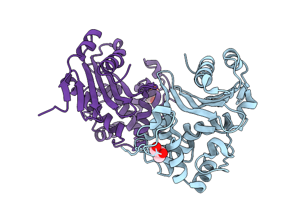 |
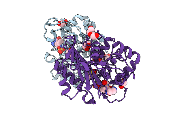
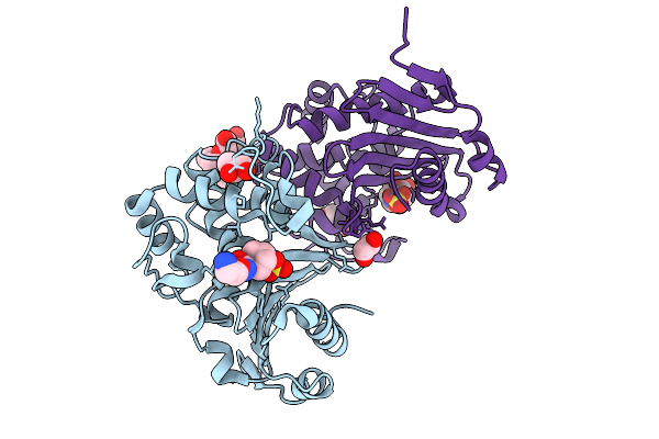
Organism: Klebsiella pneumoniae
Method: X-RAY DIFFRACTION Release Date: 2025-09-03 Classification: ANTIMICROBIAL PROTEIN Ligands: CL, GOL |
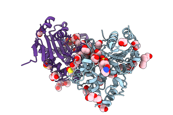 |
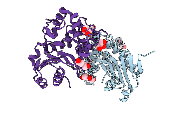
Organism: Klebsiella pneumoniae
Method: X-RAY DIFFRACTION Release Date: 2025-09-03 Classification: ANTIMICROBIAL PROTEIN Ligands: CL, PGE, PEG, EDO, OP0, DMS, GOL, PG4 |
 |
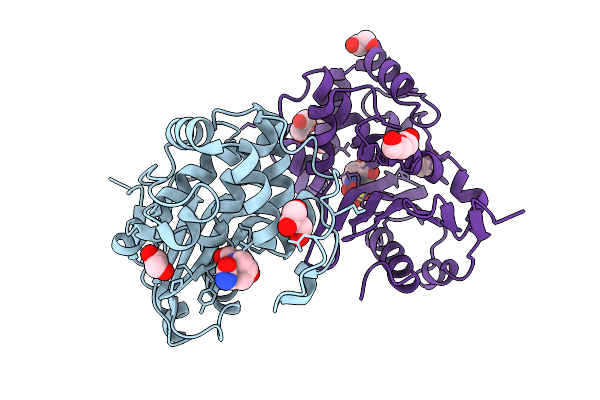
Organism: Enterobacter cloacae
Method: X-RAY DIFFRACTION Release Date: 2025-09-03 Classification: ANTIMICROBIAL PROTEIN Ligands: CL, GOL, EDO, PEG |
 |
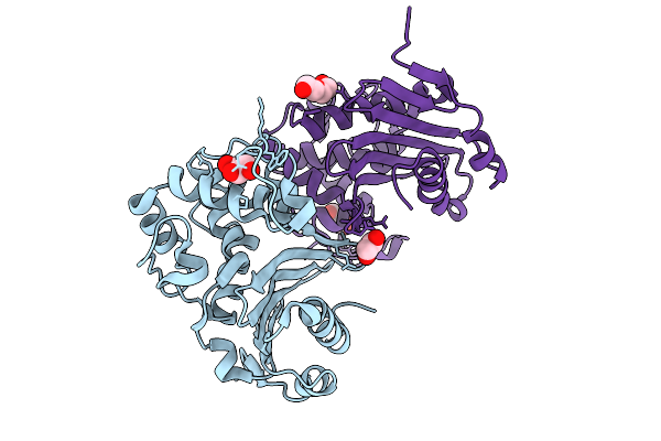
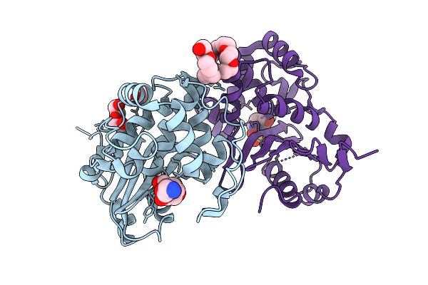
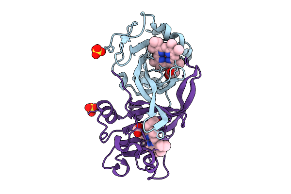
Organism: Enterobacter cloacae
Method: X-RAY DIFFRACTION Release Date: 2025-09-03 Classification: ANTIMICROBIAL PROTEIN Ligands: NXL, A1IY4, CL, EDO, 2PE |
 |
Organism: Enterobacter cloacae
Method: X-RAY DIFFRACTION Release Date: 2025-09-03 Classification: ANTIMICROBIAL PROTEIN Ligands: OP0, EDO, GOL, PEG, CL, DMS |
 |
Organism: Serratia marcescens
Method: X-RAY DIFFRACTION Release Date: 2025-09-03 Classification: ANTIMICROBIAL PROTEIN Ligands: PEG, CL, GOL, PG4, EDO |
 |
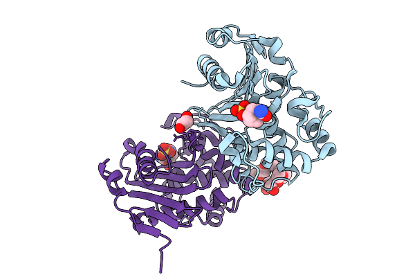
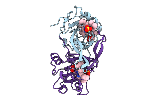
Organism: Serratia marcescens
Method: X-RAY DIFFRACTION Release Date: 2025-09-03 Classification: ANTIMICROBIAL PROTEIN Ligands: GOL, EDO, NXL, A1IY4, CL, PGE, PEG |
 |
Organism: Serratia marcescens
Method: X-RAY DIFFRACTION Release Date: 2025-09-03 Classification: ANTIMICROBIAL PROTEIN Ligands: OP0, PEG, EDO, A1IYS, CL, 2PE |
 |
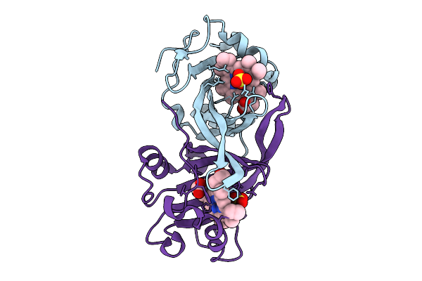
Organism: Enterobacter cloacae
Method: X-RAY DIFFRACTION Release Date: 2025-09-03 Classification: ANTIMICROBIAL PROTEIN Ligands: OP0, EDO, GOL, A1IYS, CL, 2PE |
 |
Cytochrome P460 From Methyloccocus Capsulatus In The Ferrous State (Single Crosslink From Lys)
Organism: Methylococcus capsulatus str. bath
Method: X-RAY DIFFRACTION Release Date: 2025-09-03 Classification: OXIDOREDUCTASE Ligands: SO4, HEC |
 |
Organism: Methylococcus capsulatus str. bath
Method: X-RAY DIFFRACTION Release Date: 2025-09-03 Classification: OXIDOREDUCTASE Ligands: FE, SO4, HEC |
 |
Cytochrome P460 From Methyloccocus Capsulatus (Double Crosslink From Lys), Aged
Organism: Methylococcus capsulatus str. bath
Method: X-RAY DIFFRACTION Release Date: 2025-09-03 Classification: OXIDOREDUCTASE Ligands: FE, SO4, HEC |
 |
Cytochrome P460 From Methyloccocus Capsulatus (Unclear Crosslink From Lys), Damage-Free
Organism: Methylococcus capsulatus str. bath
Method: X-RAY DIFFRACTION Release Date: 2025-09-03 Classification: OXIDOREDUCTASE Ligands: HEC, SO4 |
 |
Organism: Klebsiella pneumoniae
Method: X-RAY DIFFRACTION Resolution:1.13 Å Release Date: 2025-01-22 Classification: ANTIMICROBIAL PROTEIN Ligands: GOL, SO4 |
 |
Organism: Klebsiella pneumoniae
Method: X-RAY DIFFRACTION Resolution:1.19 Å Release Date: 2025-01-22 Classification: ANTIMICROBIAL PROTEIN Ligands: GOL, NXL, A1H3M, SO4 |
 |
Organism: Klebsiella pneumoniae
Method: X-RAY DIFFRACTION Resolution:1.03 Å Release Date: 2025-01-22 Classification: ANTIMICROBIAL PROTEIN Ligands: SO4, GOL, A1H3N, A1H3O |
 |
Organism: Klebsiella pneumoniae
Method: X-RAY DIFFRACTION Resolution:1.09 Å Release Date: 2025-01-22 Classification: ANTIMICROBIAL PROTEIN Ligands: SO4, O2F, MER |
 |
Organism: Klebsiella pneumoniae
Method: X-RAY DIFFRACTION Resolution:1.05 Å Release Date: 2024-12-25 Classification: ANTIMICROBIAL PROTEIN Ligands: GOL, SO4 |
 |
Crystal Structure Of Kemp Eliminase Hg3.17 In Complexed With 5-Cyanobenzotriazole
Organism: Thermoascus aurantiacus
Method: X-RAY DIFFRACTION Resolution:2.00 Å Release Date: 2024-09-04 Classification: LYASE Ligands: VWF, SO4 |
 |
Oxa-48_F72L. Epistasis Arises From Shifting The Rate-Limiting Step During Enzyme Evolution
Organism: Klebsiella pneumoniae
Method: X-RAY DIFFRACTION Resolution:1.97 Å Release Date: 2024-02-14 Classification: HYDROLASE Ligands: CL |