
Search Count: 23
 |
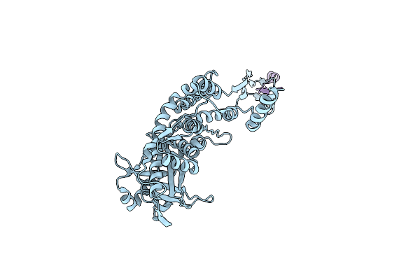
Organism: Physeter catodon
Method: X-RAY DIFFRACTION Release Date: 2025-10-15 Classification: OXIDOREDUCTASE Ligands: HEM, O |
 |
Organism: Physeter catodon
Method: X-RAY DIFFRACTION Release Date: 2025-10-15 Classification: OXIDOREDUCTASE Ligands: HEM, PEG, SO4, CL |
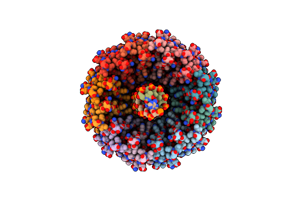 |
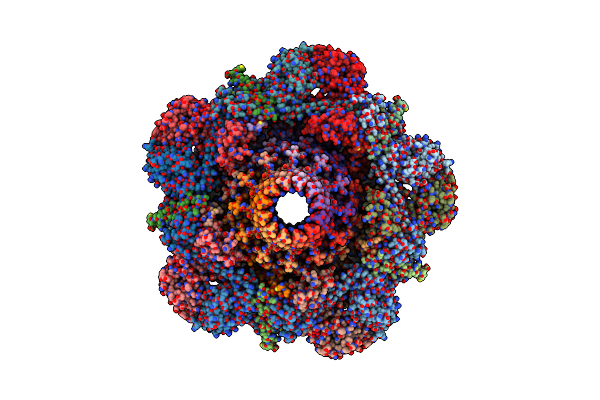
Sapi1 Neck Structure With Dna, Tail Completion Protein, And Tape Measure Protein
Organism: Staphylococcus phage 80alpha
Method: ELECTRON MICROSCOPY Release Date: 2025-06-11 Classification: VIRUS LIKE PARTICLE |
 |
Organism: Staphylococcus phage 80alpha
Method: ELECTRON MICROSCOPY Release Date: 2025-05-07 Classification: VIRUS LIKE PARTICLE |
 |
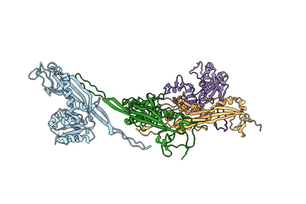
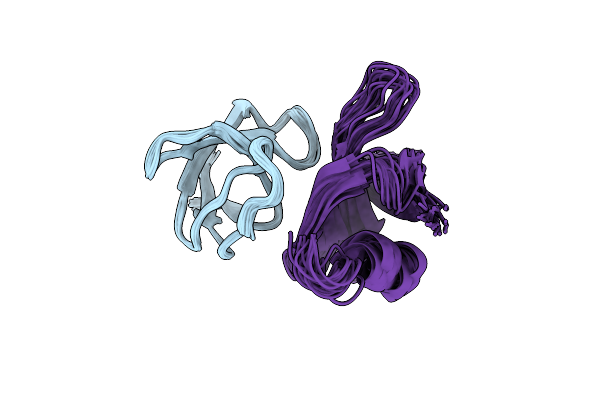
The Crystal Structure Of Human Vista Extra Cellular Domain In Complex With Fab Fragment Of Ph-Selective Anti-Vista Antibody
Organism: Homo sapiens
Method: X-RAY DIFFRACTION Resolution:2.59 Å Release Date: 2024-03-20 Classification: PROTEIN BINDING,IMMUNE SYSTEM Ligands: EDO, NAG |
 |
Organism: Staphylococcus phage 80alpha
Method: ELECTRON MICROSCOPY Release Date: 2024-01-10 Classification: STRUCTURAL PROTEIN |
 |
Organism: Dubowvirus dv80alpha
Method: ELECTRON MICROSCOPY Release Date: 2024-01-10 Classification: VIRUS LIKE PARTICLE |
 |
Organism: Dubowvirus dv80alpha
Method: ELECTRON MICROSCOPY Release Date: 2024-01-10 Classification: VIRUS LIKE PARTICLE |
 |
Organism: Dubowvirus dv80alpha
Method: ELECTRON MICROSCOPY Release Date: 2024-01-10 Classification: STRUCTURAL PROTEIN |
 |
Organism: Dubowvirus dv80alpha
Method: ELECTRON MICROSCOPY Release Date: 2024-01-10 Classification: STRUCTURAL PROTEIN |
 |
Organism: Dubowvirus dv80alpha
Method: ELECTRON MICROSCOPY Release Date: 2024-01-10 Classification: STRUCTURAL PROTEIN |
 |
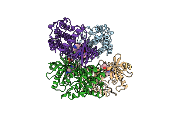
Structure Of Radical S-Adenosylmethionine Methyltransferase, Tsrm, From Kitasatospora Setae With Cobalamin And [4Fe-4S] Cluster Bound
Organism: Kitasatospora setae
Method: X-RAY DIFFRACTION Resolution:1.67 Å Release Date: 2020-12-23 Classification: TRANSFERASE Ligands: SF4, B12, EDO |
 |
Structure Of Radical S-Adenosylmethionine Methyltransferase, Tsrm, From Kitasatospora Setae With Tryptophan Substrate And Sam Analog (Aza-Sam) Bound
Organism: Kitasatospora setae
Method: X-RAY DIFFRACTION Resolution:2.19 Å Release Date: 2020-12-23 Classification: TRANSFERASE Ligands: SF4, B12, SA8, TRP |
 |
Organism: Homo sapiens
Method: X-RAY DIFFRACTION Resolution:2.19 Å Release Date: 2020-11-25 Classification: TRANSFERASE/TRANSFERASE INHIBITOR Ligands: EXF, SO4 |
 |
Crystal Structure Of Interleukin-1 Receptor-Associated Kinase 4 (Irak4-Wt) Complex With A Nicotinamide Inhibitor
Organism: Homo sapiens
Method: X-RAY DIFFRACTION Resolution:2.07 Å Release Date: 2020-06-24 Classification: TRANSFERASE/TRANSFERASE Inhibitor Ligands: R7S, SO4 |
 |
Organism: Homo sapiens, Sulfolobus solfataricus
Method: SOLUTION NMR Release Date: 2019-09-11 Classification: SIGNALING PROTEIN |
 |
Organism: Drosophila melanogaster
Method: X-RAY DIFFRACTION Resolution:1.50 Å Release Date: 2018-02-21 Classification: PROTEIN BINDING Ligands: GOL |
 |
Organism: Danio rerio, Synthetic construct
Method: X-RAY DIFFRACTION Resolution:1.70 Å Release Date: 2013-11-20 Classification: CELL CYCLE/CELL CYCLE INHIBITOR |
 |
Crystal Structure Of Tyrosine-Trna Ligase Mutant Complexed With Unnatural Amino Acid 3-O-Methyl-Tyrosine
Organism: Methanocaldococcus jannaschii
Method: X-RAY DIFFRACTION Resolution:2.00 Å Release Date: 2013-10-30 Classification: LIGASE Ligands: 3YM |
 |
Organism: Methanocaldococcus jannaschii
Method: X-RAY DIFFRACTION Resolution:2.30 Å Release Date: 2013-04-17 Classification: LIGASE Ligands: PEG |