
Search Count: 23
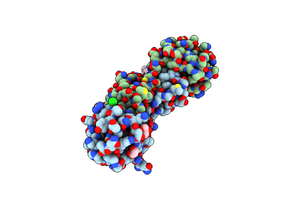 |
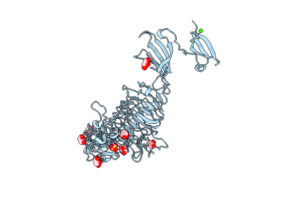
Organism: Sarcocystis muris
Method: X-RAY DIFFRACTION Resolution:1.95 Å Release Date: 2011-11-02 Classification: SUGAR BINDING PROTEIN Ligands: CL, GOL, SO4 |
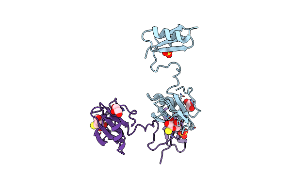 |
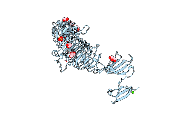
Crystal Structure Of Parasite Sarcocystis Muris Microneme Protein Sml- 2 In Complex With 1-Thio-Beta-D-Galactose (Spacegroup C2221)
Organism: Sarcocystis muris
Method: X-RAY DIFFRACTION Resolution:2.43 Å Release Date: 2011-11-02 Classification: SUGAR BINDING PROTEIN Ligands: CL, SO4, GOL, YIO |
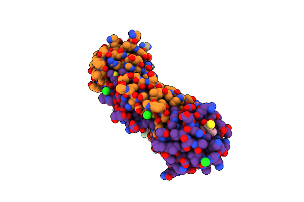 |
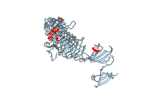
Crystal Structure Of Parasite Sarcocystis Muris Microneme Protein Sml- 2 In Complex With 1-Thio-Beta-D-Galactose (Spacegroup P212121)
Organism: Sarcocystis muris
Method: X-RAY DIFFRACTION Resolution:2.14 Å Release Date: 2011-11-02 Classification: SUGAR BINDING PROTEIN Ligands: YIO, CL, GOL |
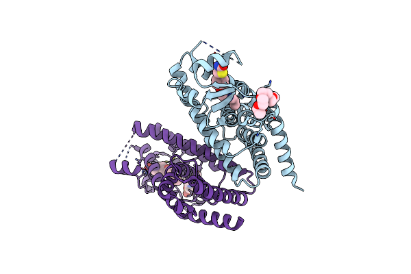 |
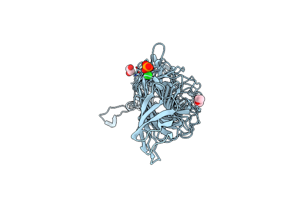
Ligand Binding Domain Of Human Ppar-Gamma In Complex With The Agonist Pioglitazone
Organism: Homo sapiens
Method: X-RAY DIFFRACTION Resolution:2.02 Å Release Date: 2011-08-10 Classification: TRANSCRIPTION Ligands: P1B, PE4, EPE |
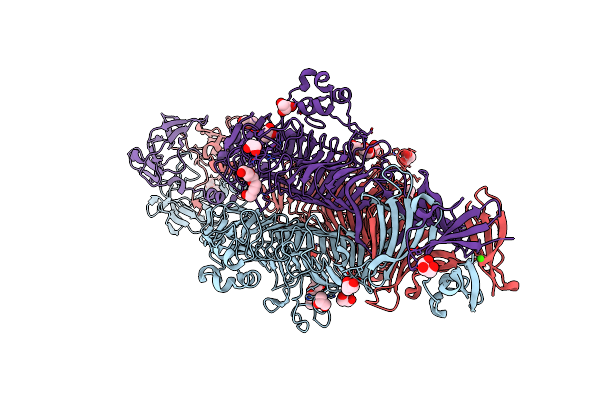 |
Organism: Enterobacteria phage p22
Method: X-RAY DIFFRACTION Resolution:1.65 Å Release Date: 2011-05-04 Classification: HYDROLASE Ligands: GOL, CA, PE4, SO4 |
 |
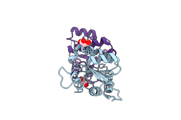
Adrenodoxin-Like Ferredoxin Etp1Fd(516-618) Of Schizosaccharomyces Pombe Mitochondria
Organism: Schizosaccharomyces pombe
Method: X-RAY DIFFRACTION Resolution:2.60 Å Release Date: 2010-08-25 Classification: ELECTRON TRANSPORT Ligands: FES |
 |
Organism: Klebsiella pneumoniae
Method: X-RAY DIFFRACTION Resolution:1.68 Å Release Date: 2010-04-28 Classification: HYDROLASE |
 |
Organism: Klebsiella pneumoniae
Method: X-RAY DIFFRACTION Resolution:2.57 Å Release Date: 2010-04-28 Classification: HYDROLASE Ligands: SO4 |
 |
Organism: Klebsiella pneumoniae
Method: X-RAY DIFFRACTION Resolution:2.57 Å Release Date: 2010-04-28 Classification: HYDROLASE Ligands: SO4 |
 |
Organism: Homo sapiens
Method: X-RAY DIFFRACTION Resolution:2.40 Å Release Date: 2009-03-03 Classification: APOPTOSIS Ligands: MLA |
 |
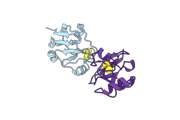
Crystal Structure Of Kora Bound To Operator Dna: Insight Into Repressor Cooperation In Rp4 Gene Regulation
Organism: Escherichia coli
Method: X-RAY DIFFRACTION Resolution:1.85 Å Release Date: 2009-02-17 Classification: TRANSCRIPTION/DNA |
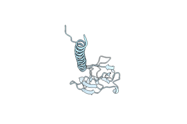 |
Headbinding Domain Of Phage P22 Tailspike C-Terminally Fused To Isoleucine Zipper Piigcn4 (Chimera I)
Organism: Enterobacteria phage p22, Saccharomyces cerevisiae
Method: X-RAY DIFFRACTION Resolution:2.05 Å Release Date: 2009-02-10 Classification: VIRAL PROTEIN |
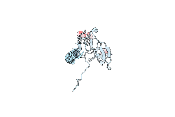 |
Mutant Y108Wdel Of The Headbinding Domain Of Phage P22 Tailspike C- Terminally Fused To Isoleucine Zipper Piigcn4 (Chimera Ii)
Organism: Enterobacteria phage p22, Saccharomyces cerevisiae
Method: X-RAY DIFFRACTION Resolution:1.80 Å Release Date: 2009-02-10 Classification: VIRAL PROTEIN Ligands: GOL |
 |
Organism: Enterobacteria phage p22
Method: X-RAY DIFFRACTION Resolution:1.50 Å Release Date: 2008-12-16 Classification: HYDROLASE Ligands: GOL, SO4, CA |
 |
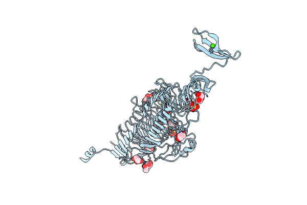
Low Temperature Structure Of P22 Tailspike Protein Fragment (109-666), Mutant V125A
Organism: Enterobacteria phage p22
Method: X-RAY DIFFRACTION Resolution:1.50 Å Release Date: 2008-12-16 Classification: HYDROLASE Ligands: GOL, SO4, CA |
 |
Low Temperature Structure Of P22 Tailspike Protein Fragment (109-666), Mutant V125L
Organism: Bacteriophage p22
Method: X-RAY DIFFRACTION Resolution:1.50 Å Release Date: 2008-12-16 Classification: HYDROLASE Ligands: GOL, SO4, CA |
 |
Low Temperature Structure Of P22 Tailspike Protein Fragment (109-666), Mutant V349L
Organism: Enterobacteria phage p22
Method: X-RAY DIFFRACTION Resolution:1.55 Å Release Date: 2008-12-16 Classification: HYDROLASE Ligands: GOL, SO4, CA |
 |
Low Temperature Structure Of P22 Tailspike Protein Fragment (109-666), Mutant V450A
Organism: Enterobacteria phage p22
Method: X-RAY DIFFRACTION Resolution:1.55 Å Release Date: 2008-12-16 Classification: HYDROLASE Ligands: GOL, SO4, CA |
 |
Organism: Bacteriophage hk620
Method: X-RAY DIFFRACTION Resolution:1.38 Å Release Date: 2008-07-01 Classification: VIRAL PROTEIN Ligands: EDO, PG0, PO4, CL |
 |
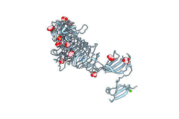
Tailspike Protein Of E.Coli Bacteriophage Hk620 In Complex With Hexasaccharide
Organism: Salmonella phage hk620
Method: X-RAY DIFFRACTION Resolution:1.59 Å Release Date: 2008-07-01 Classification: VIRAL PROTEIN Ligands: CA, CL, K, GOL |