
Search Count: 13
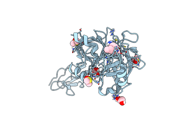 |
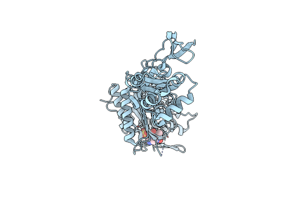
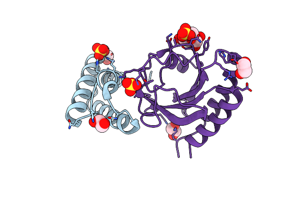
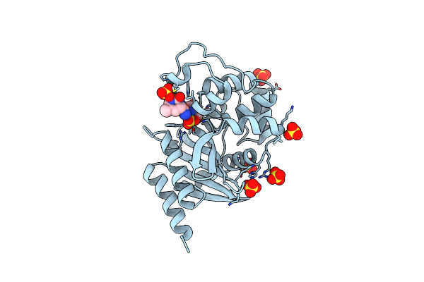
Crystal Structure Of The Catalytic Region Of Human Masp-2 With Specific Inhibitor Compound S1
Organism: Homo sapiens
Method: X-RAY DIFFRACTION Resolution:1.97 Å Release Date: 2025-04-16 Classification: HYDROLASE Ligands: A1A1X, GOL, DMS |
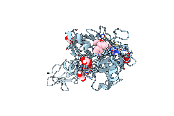 |
Crystal Structure Of The Catalytic Region Of Human Masp-2 With Specific Inhibitor Compound S2
Organism: Homo sapiens
Method: X-RAY DIFFRACTION Resolution:2.08 Å Release Date: 2025-04-16 Classification: HYDROLASE Ligands: GOL, A1A1Y |
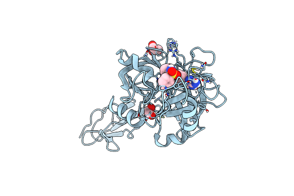 |
Crystal Structure Of The Catalytic Region Of Human Masp-2 With Specific Inhibitor Analog 20
Organism: Homo sapiens
Method: X-RAY DIFFRACTION Resolution:1.76 Å Release Date: 2025-04-16 Classification: HYDROLASE/INHIBITOR Ligands: GOL, A1A1Z |
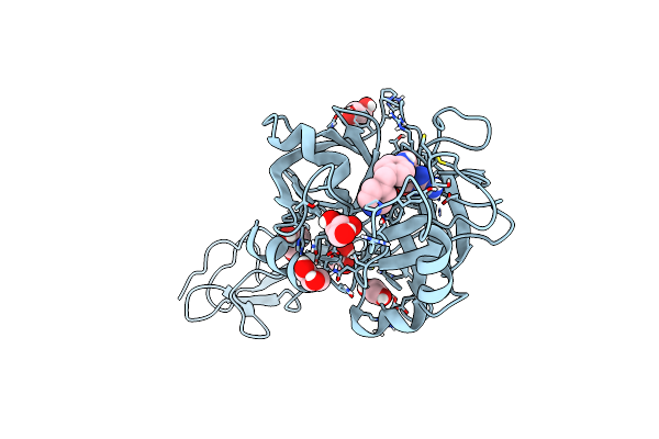 |
Crystal Structure Of The Catalytic Region Of Human Masp-2 With Specific Inhibitor Compound S3
Organism: Homo sapiens
Method: X-RAY DIFFRACTION Resolution:2.36 Å Release Date: 2025-04-16 Classification: HYDROLASE Ligands: GOL, A1A10 |
 |
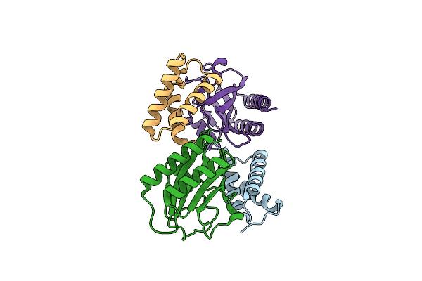
Crystal Structure Of Pseudomonas Aeruginosa Penicillin Binding Protein 3 (Pae-Pbp3) Bound To Etx0462
Organism: Pseudomonas aeruginosa
Method: X-RAY DIFFRACTION Resolution:2.20 Å Release Date: 2021-05-26 Classification: ANTIBIOTIC Ligands: CL, VMM |
 |
Crystal Structure Of P. Aeruginosa Lpxc With N-Hydroxyformamide Inhibitor 19
Organism: Pseudomonas aeruginosa (strain atcc 15692 / dsm 22644 / cip 104116 / jcm 14847 / lmg 12228 / 1c / prs 101 / pao1)
Method: X-RAY DIFFRACTION Resolution:1.90 Å Release Date: 2020-11-25 Classification: HYDROLASE Ligands: GOL, W4M, SO4, ZN |
 |
Organism: Pseudomonas aeruginosa (strain atcc 15692 / dsm 22644 / cip 104116 / jcm 14847 / lmg 12228 / 1c / prs 101 / pao1)
Method: X-RAY DIFFRACTION Resolution:2.00 Å Release Date: 2020-11-25 Classification: HYDROLASE Ligands: W4P, SO4, GOL, DMS, ZN, W8P |
 |
Organism: Enterobacteria phage t4
Method: X-RAY DIFFRACTION Resolution:2.21 Å Release Date: 2020-06-24 Classification: VIRAL PROTEIN Ligands: EDO |
 |
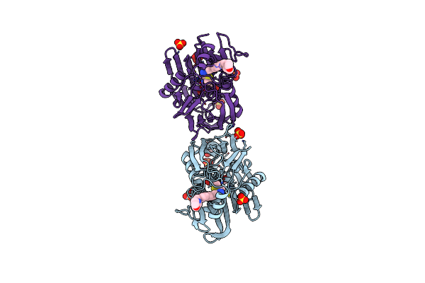
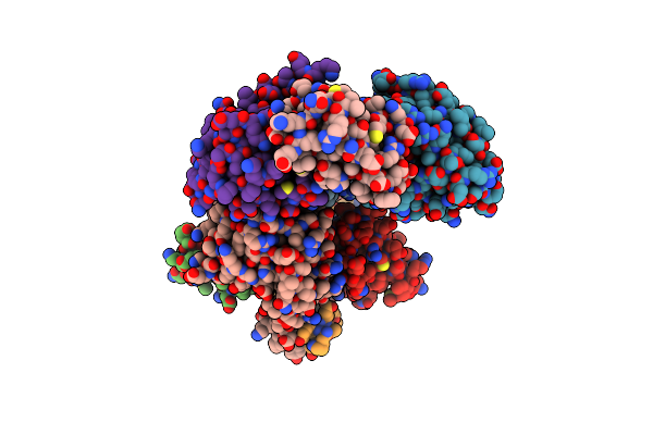
Crystal Structure Of The Complex Between Periplasmic Domains Of Antiholin Ri And Holin T From T4 Phage, In P6522
Organism: Escherichia phage ecml-134, Escherichia phage vb_ecom_nbg2
Method: X-RAY DIFFRACTION Resolution:2.20 Å Release Date: 2020-06-24 Classification: VIRAL PROTEIN Ligands: SO4, EDO, BTB, CL |
 |
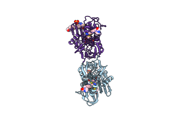
Crystal Structure Of The Complex Between Periplasmic Domains Of Antiholin Ri And Holin T From T4 Phage, In H32
Organism: Escherichia phage ecml-134, Escherichia phage vb_ecom_nbg2
Method: X-RAY DIFFRACTION Resolution:1.65 Å Release Date: 2020-06-24 Classification: VIRAL PROTEIN |
 |
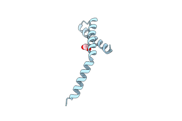
Crystal Structure Of The Complex Between Periplasmic Domains Of Antiholin Ri And Holin T From T4 Phage, In P21
Organism: Enterobacteria phage t4, Escherichia phage vb_ecom_nbg2
Method: X-RAY DIFFRACTION Resolution:2.30 Å Release Date: 2020-06-24 Classification: VIRAL PROTEIN |
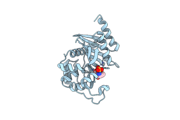 |
1.95 Ang Crystal Structure Of Oxa-24/40 Beta-Lactamase In Complex The Inhibitor Etx2514
Organism: Acinetobacter baumannii
Method: X-RAY DIFFRACTION Resolution:1.95 Å Release Date: 2019-02-27 Classification: HYDROLASE/HYDROLASE Inhibitor Ligands: JXG, CL |
 |
Diazabicyclooctenone Etx2514 Bound To Class D Beta Lactamase Oxa-24 From A. Baumannii
Organism: Acinetobacter baumannii
Method: X-RAY DIFFRACTION Resolution:1.93 Å Release Date: 2017-06-07 Classification: HYDROLASE/HYDROLASE INHIBITOR Ligands: SO4, 9CP, 9CM |