
Search Count: 24
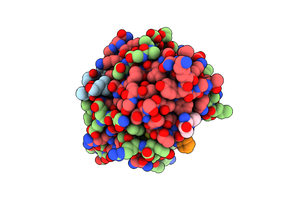 |
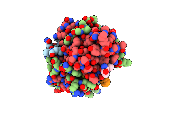
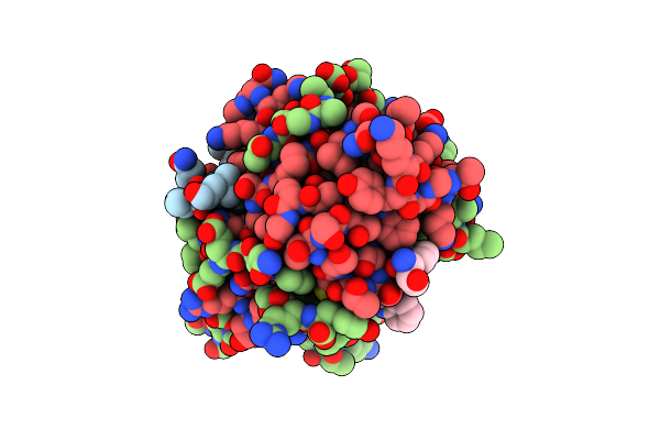
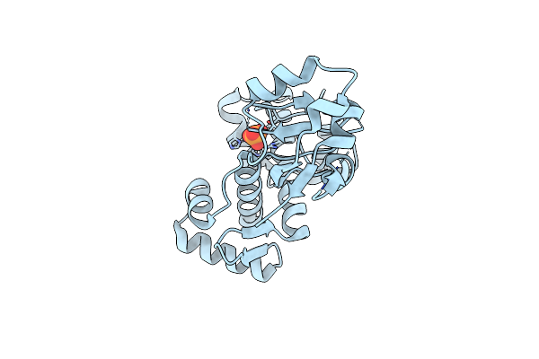
Crystal Structure Of Deuterated Gamma-Chymotrypsin At Ph 7.5, Room Temperature
Organism: Bos taurus
Method: X-RAY DIFFRACTION Resolution:1.05 Å Release Date: 2021-09-01 Classification: HYDROLASE Ligands: IOD |
 |
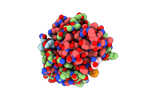
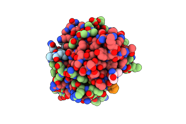
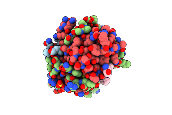
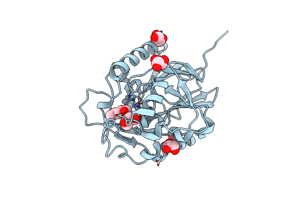
Crystal Structure Of Deuterated Gamma-Chymotrypsin At Ph 7.5, Cryo Temperature
Organism: Bos taurus
Method: X-RAY DIFFRACTION Resolution:1.00 Å Release Date: 2021-09-01 Classification: HYDROLASE Ligands: MLA, IOD |
 |
Organism: Bos taurus
Method: X-RAY DIFFRACTION Resolution:1.05 Å Release Date: 2021-09-01 Classification: HYDROLASE Ligands: IOD |
 |
Organism: Bos taurus
Method: X-RAY DIFFRACTION Resolution:1.05 Å Release Date: 2021-09-01 Classification: HYDROLASE Ligands: MLI, IOD |
 |
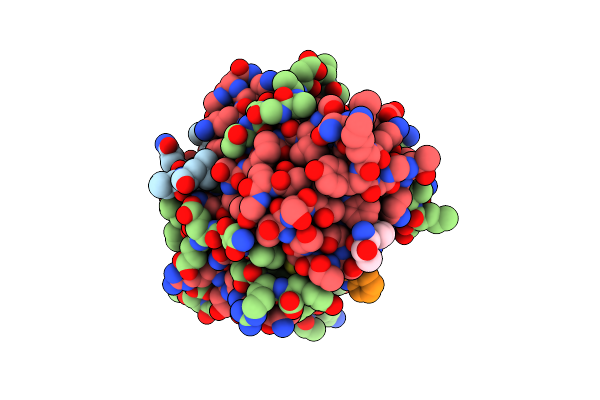
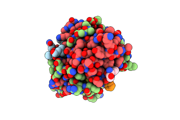
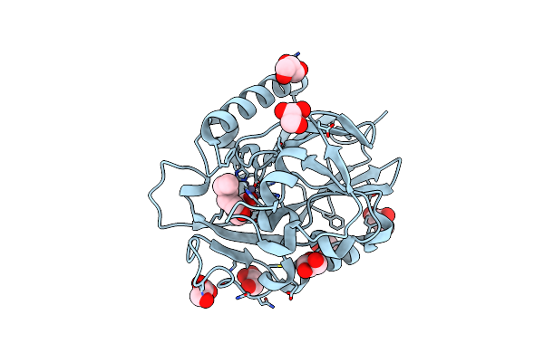
Crystal Structure Of Deuterated Gamma-Chymotrypsin At Ph 5.6, Room Temperature
Organism: Bos taurus
Method: X-RAY DIFFRACTION Resolution:1.05 Å Release Date: 2021-09-01 Classification: HYDROLASE Ligands: IOD, SO4 |
 |
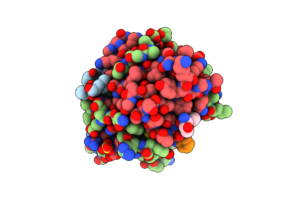
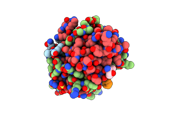
Crystal Structure Of Deuterated Gamma-Chymotrypsin At Ph 5.6, Cryo Temperature
Organism: Bos taurus
Method: X-RAY DIFFRACTION Resolution:1.10 Å Release Date: 2021-09-01 Classification: HYDROLASE Ligands: MLA, IOD |
 |
Organism: Bos taurus
Method: X-RAY DIFFRACTION Resolution:1.05 Å Release Date: 2021-09-01 Classification: HYDROLASE Ligands: IOD, SO4 |
 |
Organism: Bos taurus
Method: X-RAY DIFFRACTION Resolution:1.05 Å Release Date: 2021-09-01 Classification: HYDROLASE Ligands: MLI, IOD |
 |
Crystal Structure Of Deuterated Gamma-Chymotrypsin At Ph 9, Room Temperature
Organism: Bos taurus
Method: X-RAY DIFFRACTION Resolution:1.20 Å Release Date: 2021-09-01 Classification: HYDROLASE Ligands: IOD, SO4 |
 |
Crystal Structure Of Deuterated Gamma-Chymotrypsin At Ph 9, Cryo Temperature
Organism: Bos taurus
Method: X-RAY DIFFRACTION Resolution:1.05 Å Release Date: 2021-09-01 Classification: HYDROLASE Ligands: SO4, IOD |
 |
Organism: Bos taurus
Method: X-RAY DIFFRACTION Resolution:1.15 Å Release Date: 2021-09-01 Classification: HYDROLASE Ligands: IOD, SO4 |
 |
Organism: Bos taurus
Method: X-RAY DIFFRACTION Resolution:1.05 Å Release Date: 2021-09-01 Classification: HYDROLASE Ligands: IOD, SO4 |
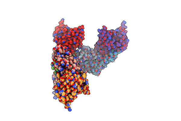 |
Organism: Homo sapiens
Method: X-RAY DIFFRACTION Resolution:3.00 Å Release Date: 2014-09-24 Classification: IMMUNE SYSTEM |
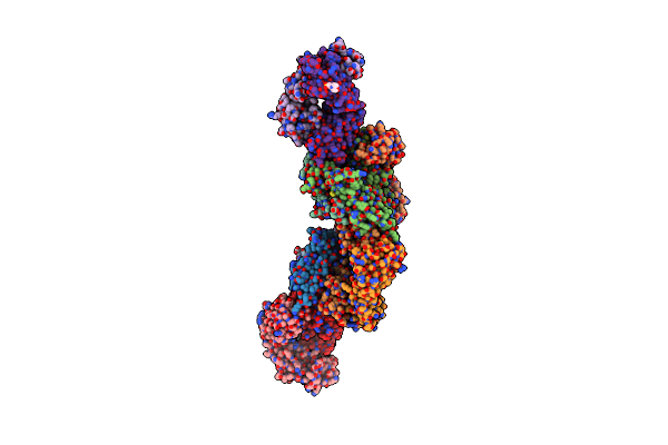 |
Organism: Homo sapiens
Method: X-RAY DIFFRACTION Resolution:2.83 Å Release Date: 2014-09-24 Classification: IMMUNE SYSTEM Ligands: MES, PE5 |
 |
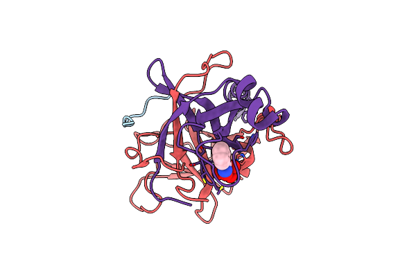
Organism: Corynebacterium diphtheriae
Method: X-RAY DIFFRACTION Resolution:1.85 Å Release Date: 2009-06-09 Classification: TRANSCRIPTION Ligands: NI, PO4 |
 |
An Ultral High Resolution Structure Of N-Acyl Homoserine Lactone Hydrolase With The Product N-Hexanoyl-L-Homoserine Bound At An Alternative Site
Organism: Bacillus thuringiensis serovar kurstaki
Method: X-RAY DIFFRACTION Resolution:0.95 Å Release Date: 2008-07-29 Classification: HYDROLASE Ligands: ZN, C6L, GOL |
 |
1.4 Angstrom Structure Of N-Acyl Homoserine Lactone Hydrolase With The Product N-Hexanoyl-L-Homoserine Bound At The Catalytic Metal Center
Organism: Bacillus thuringiensis serovar kurstaki
Method: X-RAY DIFFRACTION Resolution:1.40 Å Release Date: 2008-07-29 Classification: HYDROLASE Ligands: ZN, C6L, GOL |
 |
1.3 Angstrom Structure Of N-Acyl Homoserine Lactone Hydrolase With The Product N-Hexanoyl-L-Homocysteine Bound To The Catalytic Metal Center
Organism: Bacillus thuringiensis serovar kurstaki
Method: X-RAY DIFFRACTION Resolution:1.30 Å Release Date: 2008-07-29 Classification: HYDROLASE Ligands: ZN, CYK, GOL |
 |
X-Ray Crystallographic Characterization Of The Co(Ii)-Substituted Tris-Bound Form Of The Aminopeptidase From Aeromonas Proteolytica
Organism: Vibrio proteolyticus
Method: X-RAY DIFFRACTION Resolution:2.15 Å Release Date: 2007-06-12 Classification: HYDROLASE Ligands: CO, TRS |
 |
Crystal Structure Of A Benzohydroxamic Acid/Vanadate Complex Bound To Chymotrypsin A
Organism: Bos taurus
Method: X-RAY DIFFRACTION Resolution:1.50 Å Release Date: 2007-05-08 Classification: HYDROLASE Ligands: SO4, BVA |