
Search Count: 32
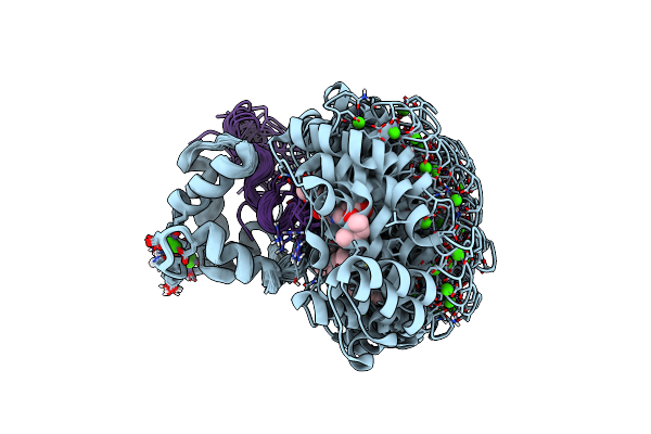 |
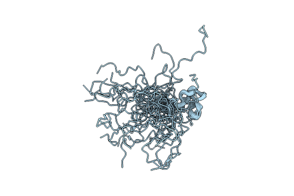
Calmodulin Extracts The Ras Family Protein Rala From Lipid Bilayers By Engagement With Two Membrane Targeting Motifs
Organism: Rattus norvegicus, Homo sapiens
Method: SOLUTION NMR Release Date: 2021-09-22 Classification: SIGNALING PROTEIN Ligands: CA, ULW |
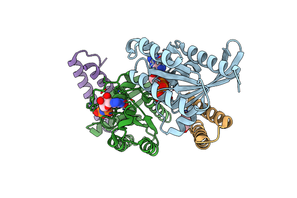 |
Crystal Structure Of The Rlip76 Ral Binding Domain Mutant (E427H/Q433L/K440R) In Complex With Ralb-Gmppnp
Organism: Homo sapiens
Method: X-RAY DIFFRACTION Resolution:1.51 Å Release Date: 2020-11-25 Classification: PROTEIN BINDING Ligands: GNP, MG, GOL |
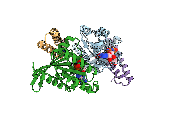 |
Crystal Structure Of The Rlip76 Ral Binding Domain Mutant (E427S/L429M/Q433L/K440R) In Complex With Ralb-Gmppnp
Organism: Homo sapiens
Method: X-RAY DIFFRACTION Resolution:1.48 Å Release Date: 2020-11-25 Classification: PROTEIN BINDING Ligands: GNP, MG, GOL |
 |
Structure Of Peptide P7, Which Binds Cdc42 And Inhibits Effector Interactions.
Organism: Synthetic construct
Method: SOLUTION NMR Release Date: 2020-01-29 Classification: DE NOVO PROTEIN |
 |
Organism: Trypanosoma brucei brucei
Method: X-RAY DIFFRACTION Resolution:1.65 Å Release Date: 2017-09-20 Classification: IMMUNE SYSTEM Ligands: GOL, MES |
 |
Organism: Trypanosoma brucei brucei
Method: SOLUTION NMR Release Date: 2017-09-20 Classification: MEMBRANE PROTEIN |
 |
Organism: Xenopus (silurana) tropicalis
Method: SOLUTION NMR Release Date: 2016-05-04 Classification: PROTEIN BINDING |
 |
Organism: Rattus norvegicus
Method: SOLID-STATE NMR, ELECTRON MICROSCOPY Release Date: 2013-12-04 Classification: PROTEIN FIBRIL |
 |
Organism: Rattus norvegicus
Method: SOLID-STATE NMR, ELECTRON MICROSCOPY Release Date: 2013-12-04 Classification: PROTEIN FIBRIL |
 |
Organism: Homo sapiens
Method: SOLUTION NMR Release Date: 2013-12-04 Classification: PROTEIN BINDING |
 |
Organism: Rattus norvegicus
Method: SOLID-STATE NMR, ELECTRON MICROSCOPY Release Date: 2013-12-04 Classification: PROTEIN FIBRIL |
 |
Organism: Homo sapiens
Method: SOLID-STATE NMR Release Date: 2013-07-17 Classification: PROTEIN FIBRIL |
 |
Organism: Homo sapiens
Method: SOLUTION NMR Release Date: 2010-09-01 Classification: TRANSPORT PROTEIN |
 |
Organism: Homo sapiens
Method: SOLUTION NMR Release Date: 2010-09-01 Classification: TRANSPORT PROTEIN, PROTEIN BINDING Ligands: GNP, MG |
 |
Organism: Natronomonas pharaonis
Method: SOLUTION NMR Release Date: 2010-06-02 Classification: MEMBRANE PROTEIN Ligands: RET |
 |
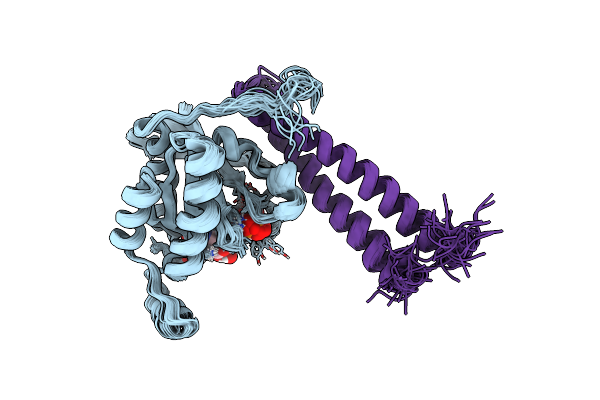
Solution Structure And Dynamics Of The Small Gtpase Ralb In Its Active Conformation: Significance For Effector Protein Binding
Organism: Homo sapiens
Method: SOLUTION NMR Release Date: 2009-02-17 Classification: SIGNALING PROTEIN Ligands: GNP, MG |
 |
Organism: Homo sapiens
Method: SOLUTION NMR Release Date: 2008-11-11 Classification: PROTEIN BINDING |
 |
Structure Of A Glycosylphosphatidylinositol-Anchored Domain From A Trypanosome Variant Surface Glycoprotein
Organism: Trypanosoma
Method: SOLUTION NMR Release Date: 2007-11-13 Classification: Membrane Protein, Immune System |
 |
Structure Of A Glycosylphosphatidylinositol-Anchored Domain From A Trypanosome Variant Surface Glycoprotein
Organism: Trypanosoma brucei brucei
Method: SOLUTION NMR Release Date: 2007-11-13 Classification: Membrane Protein, Immune System |
 |
Organism: Homo sapiens
Method: SOLUTION NMR Release Date: 2007-11-13 Classification: MEMBRANE PROTEIN/TRANSFERASE Ligands: MG, GCP |