
Search Count: 16
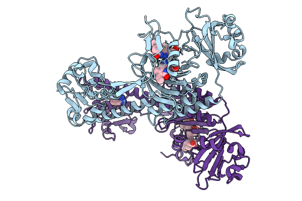 |
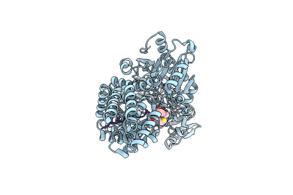
Photoactivation In Bacteriophytochromes, Reference (Dark) Structure For The 3 Ps Time Point
Organism: Stigmatella aurantiaca
Method: X-RAY DIFFRACTION Resolution:2.30 Å Release Date: 2025-10-08 Classification: SIGNALING PROTEIN Ligands: 3Q8, BEN |
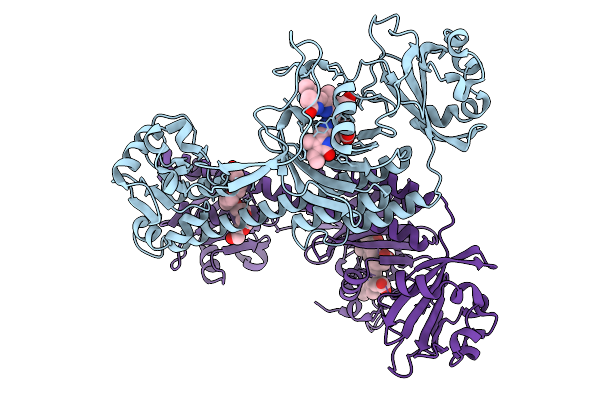 |
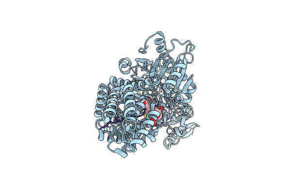
Photoactivation In Bacteriophytochrome, High Resolution Cryo Structure In The Dark.
Organism: Stigmatella aurantiaca
Method: X-RAY DIFFRACTION Resolution:1.40 Å Release Date: 2025-10-08 Classification: SIGNALING PROTEIN Ligands: EL5, P33 |
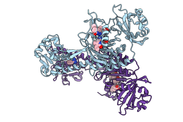 |
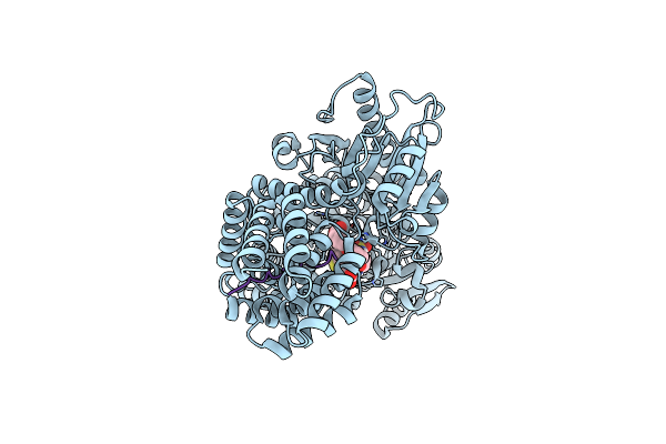
Photoactivation In Bacteriophytochromes, Reference (Dark) Structure For The 100 Ps Time Point
Organism: Stigmatella aurantiaca
Method: X-RAY DIFFRACTION Resolution:1.93 Å Release Date: 2025-10-08 Classification: SIGNALING PROTEIN Ligands: EL5, BEN |
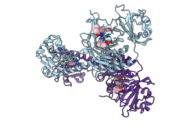 |
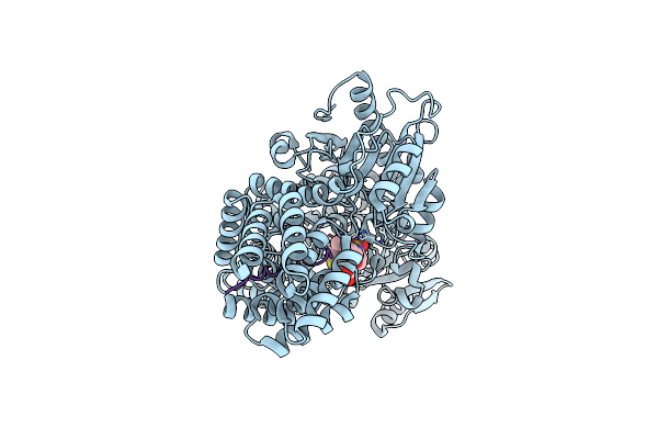
Organism: Stigmatella aurantiaca
Method: X-RAY DIFFRACTION Resolution:2.30 Å Release Date: 2025-10-08 Classification: SIGNALING PROTEIN Ligands: BLA, BEN |
 |
Organism: Stigmatella aurantiaca
Method: X-RAY DIFFRACTION Resolution:2.30 Å Release Date: 2025-10-08 Classification: SIGNALING PROTEIN Ligands: BLA, BEN |
 |
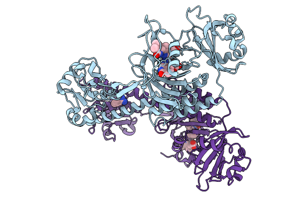
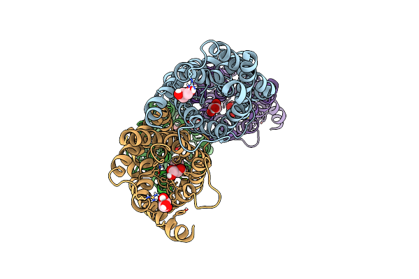
Structure Of Xenon-Derivatized Methyl-Coenzyme M Reductase From Methanothermobacter Marburgensis
Organism: Methanothermobacter marburgensis str. marburg
Method: X-RAY DIFFRACTION Resolution:2.50 Å Release Date: 2022-04-06 Classification: TRANSFERASE Ligands: F43, MG, ACT, TP7, COM, NA, XE |
 |
Xfel Serial Crystallography Reveals The Room Temperature Structure Of Methyl-Coenzyme M Reductase
Organism: Methanothermobacter marburgensis str. marburg
Method: X-RAY DIFFRACTION Resolution:1.90 Å Release Date: 2022-03-02 Classification: TRANSFERASE Ligands: MG, F43, TP7, COM, ACT, EDO |
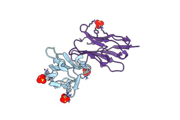 |
Organism: Synthetic construct
Method: X-RAY DIFFRACTION Resolution:2.05 Å Release Date: 2020-11-25 Classification: IMMUNE SYSTEM Ligands: SO4, CL |
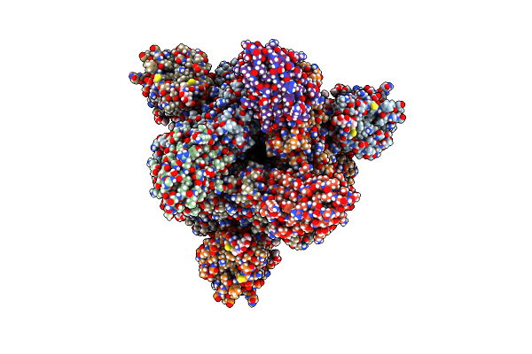 |
Organism: Severe acute respiratory syndrome coronavirus 2, Synthetic construct
Method: ELECTRON MICROSCOPY Release Date: 2020-11-11 Classification: VIRAL PROTEIN/IMMUNE SYSTEM Ligands: NAG |
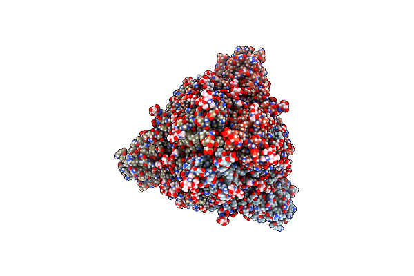 |
Organism: Severe acute respiratory syndrome coronavirus 2, Synthetic construct
Method: ELECTRON MICROSCOPY Release Date: 2020-11-11 Classification: VIRAL PROTEIN/IMMUNE SYSTEM Ligands: NAG |
 |
Organism: Homo sapiens
Method: X-RAY DIFFRACTION Resolution:4.00 Å Release Date: 2019-11-13 Classification: STRUCTURAL PROTEIN Ligands: GOL |
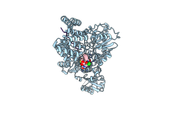 |
Crystal Structure Of Human O-Glcnac Transferase Bound To A Peptide From Hcf-1 Pro-Repeat 2 (11-26) And Inhibitor 4A
Organism: Homo sapiens
Method: X-RAY DIFFRACTION Resolution:2.75 Å Release Date: 2018-10-17 Classification: transferase/transferase inhibitor Ligands: JA4 |
 |
Crystal Structure Of Human O-Glcnac Transferase Bound To A Peptide From Hcf-1 Pro-Repeat 2 (11-26) And Inhibitor Ent-1A
Organism: Homo sapiens
Method: X-RAY DIFFRACTION Resolution:2.10 Å Release Date: 2018-10-17 Classification: transferase/transferase inhibitor Ligands: J9S |
 |
Crystal Structure Of Human O-Glcnac Transferase Bound To A Peptide From Hcf-1 Pro-Repeat 2 (11-26) And Inhibitor 2A
Organism: Homo sapiens
Method: X-RAY DIFFRACTION Resolution:2.00 Å Release Date: 2018-10-17 Classification: transferase/transferase inhibitor Ligands: JAJ |
 |
Crystal Structure Of Human O-Glcnac Transferase Bound To A Peptide From Hcf-1 Pro-Repeat 2 (11-26) And Inhibitor 3A
Organism: Homo sapiens
Method: X-RAY DIFFRACTION Resolution:2.00 Å Release Date: 2018-10-17 Classification: transferase/transferase inhibitor Ligands: JA7 |
 |
Crystal Structure Of Human O-Glcnac Transferase Bound To A Peptide From Hcf-1 Pro-Repeat 2 (11-26) And Inhibitor 1A
Organism: Homo sapiens
Method: X-RAY DIFFRACTION Resolution:2.00 Å Release Date: 2018-10-17 Classification: transferase/transferase inhibitor Ligands: J9V |