
Search Count: 15
 |
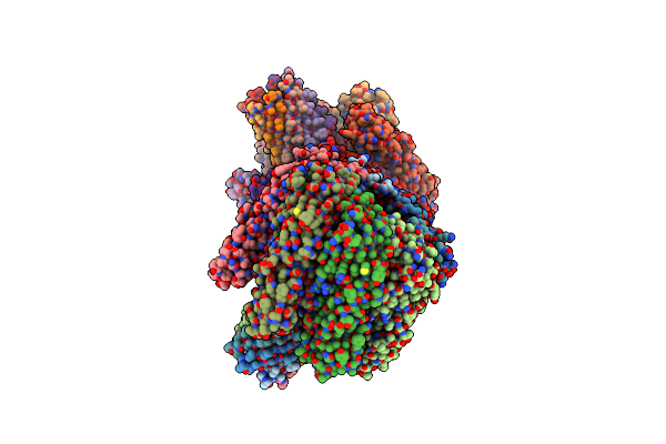
Organism: Clostridium perfringens b
Method: ELECTRON MICROSCOPY Release Date: 2019-06-19 Classification: TOXIN |
 |
Structure Of Clostridium Perfringens Enterotoxin With A Peptide Derived From A Modified Version Of Ecl-2 Of Claudin 2
Organism: Clostridium perfringens, Mus musculus
Method: X-RAY DIFFRACTION Resolution:3.38 Å Release Date: 2014-07-16 Classification: Toxin/CELL Adhesion |
 |
Clostridium Perfringens Enterotoxin, D48A Mutation And N-Terminal 37 Residues Deleted
Organism: Clostridium perfringens
Method: X-RAY DIFFRACTION Resolution:1.90 Å Release Date: 2014-01-29 Classification: TOXIN Ligands: P6G |
 |
Clostridium Perfringens Enterotoxin With The N-Terminal 37 Residues Deleted
Organism: Clostridium perfringens
Method: X-RAY DIFFRACTION Resolution:1.90 Å Release Date: 2014-01-29 Classification: TOXIN Ligands: P6G |
 |
Clostridium Perfringens Epsilon Toxin Mutant H149A Bound To Octyl Glucoside
Organism: Clostridium perfringens d
Method: X-RAY DIFFRACTION Resolution:2.40 Å Release Date: 2013-03-27 Classification: TOXIN Ligands: BOG, PO4 |
 |
Crystal Structure Of The Clostridium Perfringens Netb Toxin In The Membrane Inserted Form
Organism: Clostridium perfringens
Method: X-RAY DIFFRACTION Resolution:3.90 Å Release Date: 2012-12-26 Classification: TOXIN |
 |
Clostridium Perfringens Epsilon Toxin Shows Structural Similarity With The Pore Forming Toxin Aerolysin
Organism: Clostridium perfringens
Method: X-RAY DIFFRACTION Resolution:2.60 Å Release Date: 2004-08-05 Classification: TOXIN Ligands: U1 |
 |
Crystal Structure Analysis Of Clostridium Perfringens Alpha-Toxin Isolated From Avian Strain Swcp
Organism: Clostridium perfringens
Method: X-RAY DIFFRACTION Resolution:2.40 Å Release Date: 2002-06-19 Classification: TOXIN Ligands: ZN |
 |
Organism: Clostridium perfringens
Method: X-RAY DIFFRACTION Resolution:1.90 Å Release Date: 1999-05-04 Classification: HYDROLASE Ligands: ZN, CD |
 |
Close Packing Of An Oligomeric Eye Lens Beta-Crystallin Induces Loss Of Symmetry And Ordering Of Sequence Extensions
Organism: Bos taurus
Method: X-RAY DIFFRACTION Resolution:3.30 Å Release Date: 1994-12-20 Classification: EYE LENS PROTEIN |
 |
Organism: Bos taurus
Method: X-RAY DIFFRACTION Resolution:1.50 Å Release Date: 1994-01-31 Classification: HYDROLASE(ENDORIBONUCLEASE) Ligands: SO4, 5GP |
 |
Organism: Bos taurus
Method: X-RAY DIFFRACTION Resolution:1.50 Å Release Date: 1994-01-31 Classification: HYDROLASE(ENDORIBONUCLEASE) Ligands: SO4, DGP |
 |
Structure Of The Bovine Eye Lens Protein Gamma-B (Gamma-Ii)-Crystallin At 1.47 Angstroms
Organism: Bos taurus
Method: X-RAY DIFFRACTION Resolution:1.47 Å Release Date: 1993-10-31 Classification: EYE LENS PROTEIN |
 |
Segmented Anisotropic Refinement Of Bovine Ribonuclease A By The Application Of The Rigid-Body Tls Model
Organism: Bos taurus
Method: X-RAY DIFFRACTION Resolution:1.45 Å Release Date: 1991-10-31 Classification: HYDROLASE (NUCLEIC ACID,RNA) Ligands: SO4 |
 |
Organism: Bos taurus
Method: X-RAY DIFFRACTION, NEUTRON DIFFRACTION Resolution:2.0 Å, 2.0 Å Release Date: 1985-07-17 Classification: Hydrolase/RNA Ligands: PO4, DOD |