
Search Count: 26
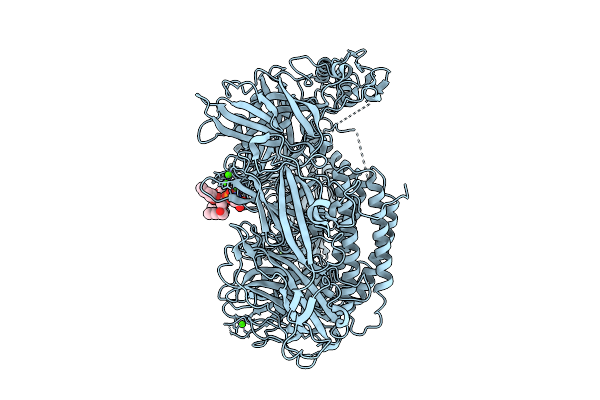 |
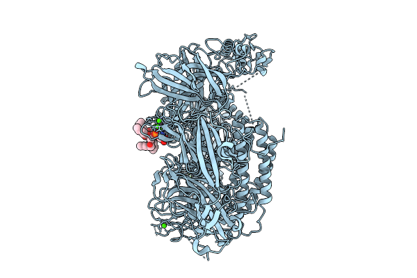
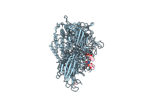
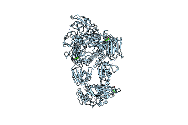
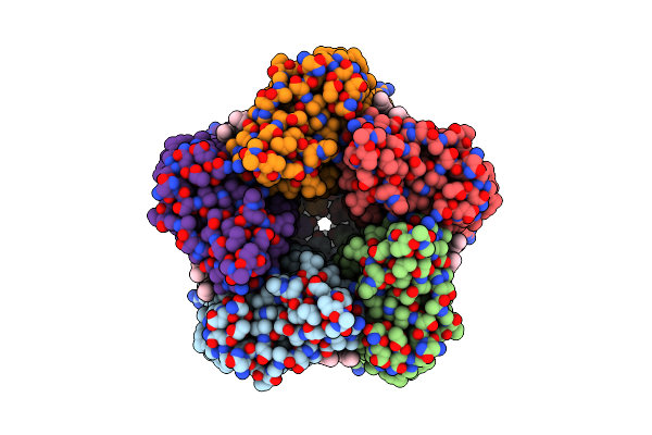
Mouse Otoferlin (216-1931) In Complex With An Msp2N2 Lipid Nanodisc (30 Mol% Dops, 10 Mol% Pi(4,5)P2)
Organism: Mus musculus
Method: ELECTRON MICROSCOPY Release Date: 2025-11-12 Classification: MEMBRANE PROTEIN Ligands: CA, PSF |
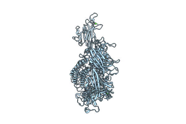 |
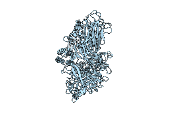
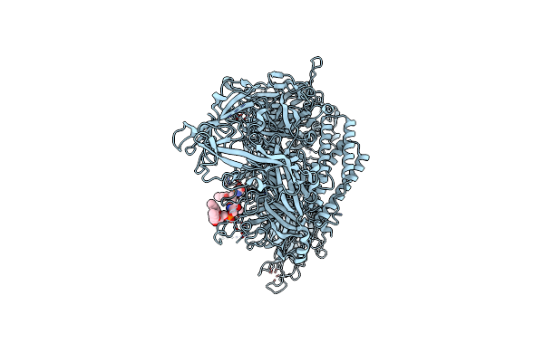
Mouse Otoferlin (216-1931) In The Lipid-Free, Ca2+-Bound State, "Open" Conformation (Class 2)
Organism: Mus musculus
Method: ELECTRON MICROSCOPY Release Date: 2025-11-05 Classification: MEMBRANE PROTEIN Ligands: CA |
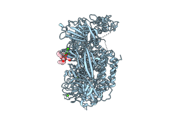 |
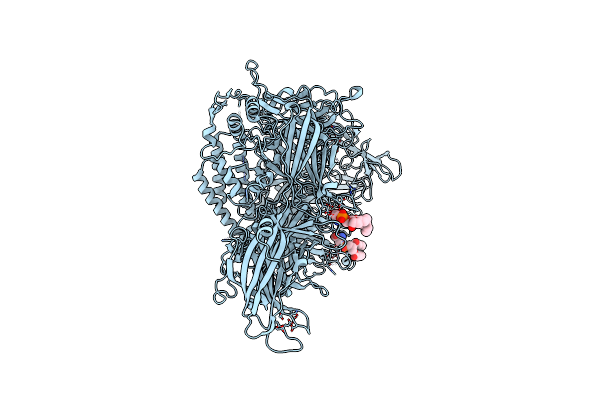
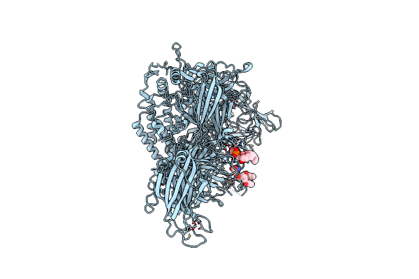
Mouse Otoferlin (216-1931) In Complex With A Lipid Nanodisc (Comprising 25% Ps And 5% Pip2)
Organism: Mus musculus
Method: ELECTRON MICROSCOPY Release Date: 2025-11-05 Classification: MEMBRANE PROTEIN Ligands: CA, PSF |
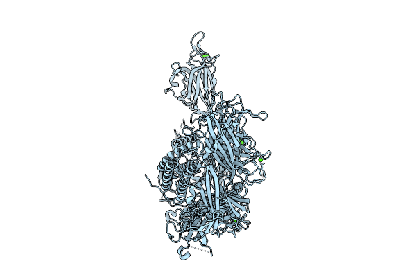 |
Mouse Otoferlin (216-1931) In The Lipid-Free Ca2+-Bound State, "Open" Conformation (Class 1)
Organism: Mus musculus
Method: ELECTRON MICROSCOPY Release Date: 2025-11-05 Classification: MEMBRANE PROTEIN Ligands: CA |
 |
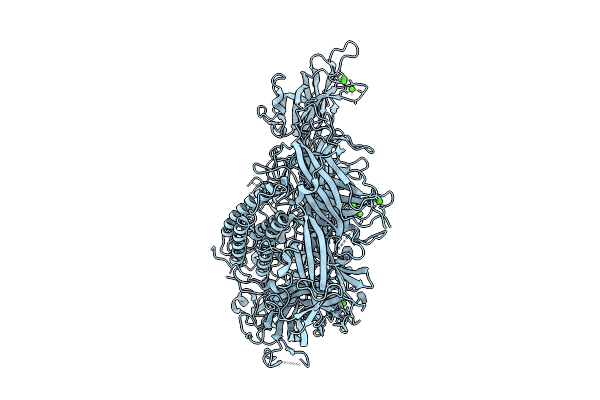
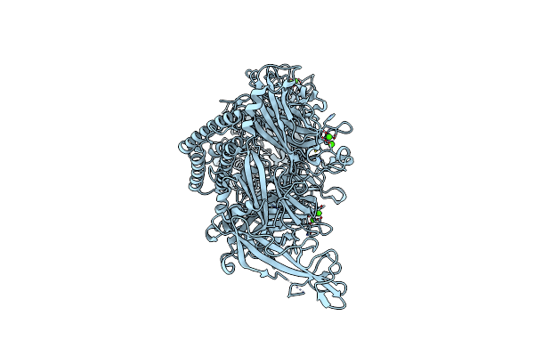
Mouse Otoferlin (Residues 216-1931) In The Lipid-Bound State (Merged Datasets)
Organism: Mus musculus
Method: ELECTRON MICROSCOPY Release Date: 2025-11-05 Classification: MEMBRANE PROTEIN Ligands: CA, PSF |
 |
Mouse Otoferlin (216-1931) In The Lipid-Free, Ca2+-Free State ("Loose" Conformation)
Organism: Mus musculus
Method: ELECTRON MICROSCOPY Release Date: 2025-11-05 Classification: MEMBRANE PROTEIN |
 |
Mouse Otoferlin (216-1931) In The Lipid-Free Ca2+-Bound State, "Closed-Like" Conformation
Organism: Mus musculus
Method: ELECTRON MICROSCOPY Release Date: 2025-11-05 Classification: MEMBRANE PROTEIN Ligands: CA |
 |
Cryo-Em Structure Of Lipid-Bound Human Myoferlin (25 Mol% Dops/5 Mol% Pi(4,5)P2 Nanodisc)
Organism: Homo sapiens
Method: ELECTRON MICROSCOPY Release Date: 2025-06-04 Classification: MEMBRANE PROTEIN Ligands: PSF, CA |
 |
Human Myoferlin (1-1997) In Complex With An Msp2N2 Lipid Nanodisc (15 Mol% Dops, 5 Mol% Cholesterol)
Organism: Homo sapiens
Method: ELECTRON MICROSCOPY Release Date: 2025-06-04 Classification: MEMBRANE PROTEIN Ligands: PSF, CA |
 |
Human Myoferlin (1-1997) In Complex With An Msp2N2 Lipid Nanodisc (15 Mol% Dops, 2 Mol% Pi(4,5)P2)
Organism: Homo sapiens
Method: ELECTRON MICROSCOPY Release Date: 2025-06-04 Classification: MEMBRANE PROTEIN Ligands: PSF, CA |
 |
Human Myoferlin (1-1997) In Complex With An Msp2N2 Lipid Nanodisc (25 Mol% Dops, 5 Mol% Pi(4,5)P2 And 5 Mol% Cholesterol)
Organism: Homo sapiens
Method: ELECTRON MICROSCOPY Release Date: 2025-06-04 Classification: MEMBRANE PROTEIN Ligands: PSF, CA |
 |
Organism: Homo sapiens
Method: ELECTRON MICROSCOPY Release Date: 2025-06-04 Classification: MEMBRANE PROTEIN Ligands: CA |
 |
Organism: Homo sapiens
Method: ELECTRON MICROSCOPY Release Date: 2025-06-04 Classification: MEMBRANE PROTEIN Ligands: CA |
 |
Organism: Cryobacterium levicorallinum
Method: ELECTRON MICROSCOPY Release Date: 2025-05-14 Classification: MEMBRANE PROTEIN Ligands: LFA, RET |
 |
Organism: Cryobacterium levicorallinum
Method: ELECTRON MICROSCOPY Release Date: 2025-05-14 Classification: MEMBRANE PROTEIN Ligands: LFA, RET |
 |
Organism: Cryobacterium levicorallinum
Method: ELECTRON MICROSCOPY Release Date: 2025-05-14 Classification: MEMBRANE PROTEIN Ligands: LMT, LFA, RET |
 |
Cryo-Em Structure Of The Microbial Rhodopsin Cryor1 At Ph 10.5 In Detergent In The Ground State
Organism: Cryobacterium levicorallinum
Method: ELECTRON MICROSCOPY Release Date: 2025-05-14 Classification: MEMBRANE PROTEIN Ligands: LMT, RET, LFA |
 |
Cryo-Em Structure Of The Microbial Rhodopsin Cryor1 At Ph 10.5 In Detergent In The M State
Organism: Cryobacterium levicorallinum
Method: ELECTRON MICROSCOPY Release Date: 2025-05-14 Classification: MEMBRANE PROTEIN Ligands: LMT, RET |
 |
Organism: Subtercola endophyticus
Method: ELECTRON MICROSCOPY Release Date: 2025-05-14 Classification: MEMBRANE PROTEIN Ligands: LFA, RET |
 |
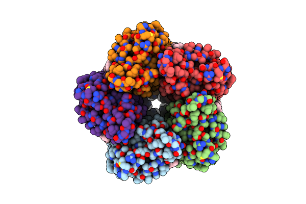
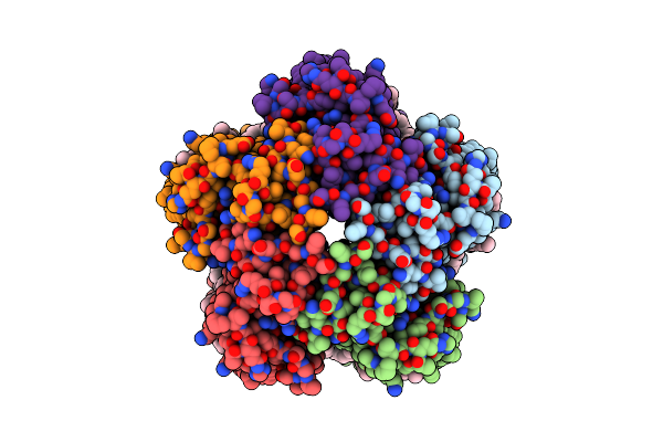
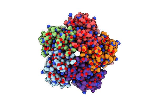
Organism: Bacillus sp. (in: firmicutes)
Method: ELECTRON MICROSCOPY Release Date: 2024-08-07 Classification: METAL TRANSPORT |