
Search Count: 26
All
Selected
 |
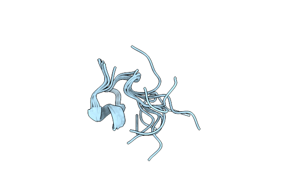
Organism: Bos taurus
Method: X-RAY DIFFRACTION Resolution:1.45 Å Release Date: 2017-05-03 Classification: HORMONE |
 |
Organism: Saccharomyces cerevisiae
Method: X-RAY DIFFRACTION Resolution:2.40 Å Release Date: 2011-02-16 Classification: hydrolase/hydrolase inhibitor Ligands: MES |
 |
Organism: Saccharomyces cerevisiae
Method: X-RAY DIFFRACTION Resolution:2.50 Å Release Date: 2011-02-16 Classification: hydrolase/hydrolase inhibitor Ligands: MES |
 |
Organism: Saccharomyces cerevisiae
Method: X-RAY DIFFRACTION Resolution:2.70 Å Release Date: 2011-02-16 Classification: hydrolase/hydrolase inhibitor |
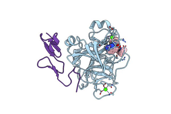 |
Factor Xa In Complex With (R)-2-(1-Adamantylcarbamoylamino)-3-(3-Carbamidoyl-Phenyl)-N-Phenethyl-Propionic Acid Amide
Organism: Homo sapiens
Method: X-RAY DIFFRACTION Resolution:2.22 Å Release Date: 2010-04-07 Classification: HYDROLASE Ligands: CA, RUP |
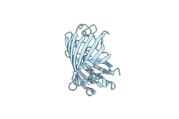 |
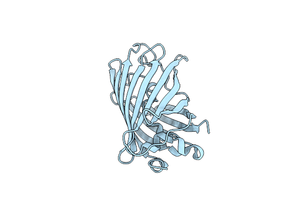
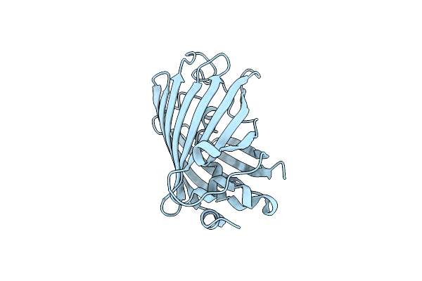
The Chemical Control Of Protein Folding: Engineering A Superfolder Green Fluorescent Protein
Organism: Aequorea victoria
Method: X-RAY DIFFRACTION Resolution:2.10 Å Release Date: 2007-06-19 Classification: LUMINESCENT PROTEIN |
 |
Organism: Saccharomyces cerevisiae
Method: X-RAY DIFFRACTION Resolution:2.81 Å Release Date: 2006-07-11 Classification: HYDROLASE Ligands: BIQ |
 |
 |
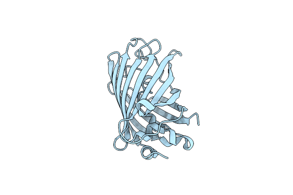
Crystallographic Evidence For Isomeric Chromophores In 3-Fluorotyrosyl-Green Fluorescent Protein
Organism: Aequorea victoria
Method: X-RAY DIFFRACTION Resolution:2.10 Å Release Date: 2004-06-08 Classification: LUMINESCENT PROTEIN |
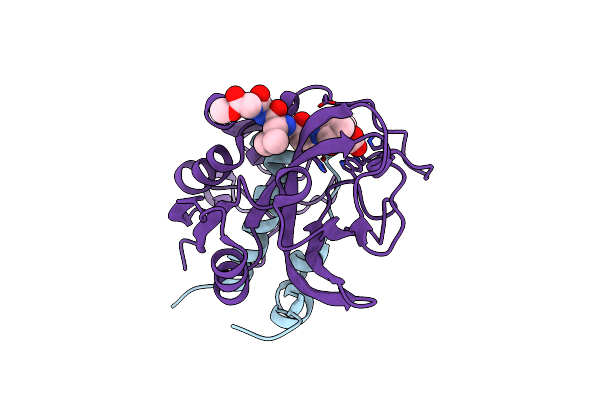 |
Crystal Structure Of Ns-134 In Complex With Bovine Cathepsin B: A Two Headed Epoxysuccinyl Inhibitor Extends Along The Whole Active Site Cleft
Organism: Bos taurus
Method: X-RAY DIFFRACTION Resolution:2.20 Å Release Date: 2004-05-04 Classification: HYDROLASE/HYDROLASE INHIBITOR Ligands: EP2 |
 |
 |
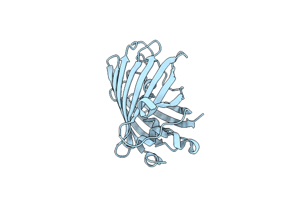
Expansion Of The Genetic Code Enables Design Of A Novel "Gold" Class Of Green Fluorescent Proteins
Organism: Cfp marker plasmid pwm1009
Method: X-RAY DIFFRACTION Resolution:1.15 Å Release Date: 2003-12-02 Classification: LUMINESCENT PROTEIN |
 |
Expansion Of The Genetic Code Enables Design Of A Novel "Gold" Class Of Green Fluorescent Proteins
Organism: Cfp marker plasmid pwm1009
Method: X-RAY DIFFRACTION Resolution:1.15 Å Release Date: 2003-12-02 Classification: LUMINESCENT PROTEIN |
 |
Expansion Of The Genetic Code Enables Design Of A Novel "Gold" Class Of Green Fluorescent Proteins
Organism: Cfp marker plasmid pwm1009
Method: X-RAY DIFFRACTION Resolution:1.69 Å Release Date: 2003-12-02 Classification: LUMINESCENT PROTEIN |
 |
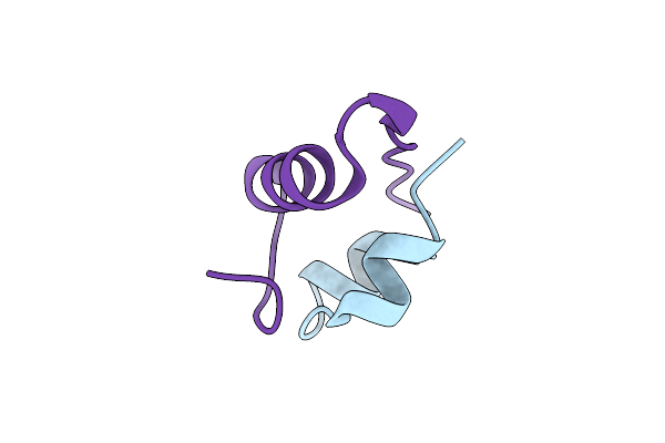
Organism: Helleborus purpurascens
Method: SOLUTION NMR Release Date: 2003-03-11 Classification: TOXIN |
 |
Tricorn Protease In Complex With Tetrapeptide Chloromethyl Ketone Derivative
Organism: Thermoplasma acidophilum, Synthetic construct
Method: X-RAY DIFFRACTION Resolution:2.80 Å Release Date: 2002-12-30 Classification: HYDROLASE Ligands: D10 |
 |
Tricorn Protease In Complex With A Tridecapeptide Chloromethyl Ketone Derivative
Organism: Thermoplasma acidophilum, Synthetic construct
Method: X-RAY DIFFRACTION Resolution:2.60 Å Release Date: 2002-12-30 Classification: HYDROLASE |
 |
Organism: Thermoplasma acidophilum
Method: X-RAY DIFFRACTION Resolution:2.70 Å Release Date: 2002-12-30 Classification: HYDROLASE Ligands: DKT |
 |
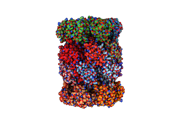
Organism: Escherichia coli
Method: X-RAY DIFFRACTION Resolution:2.06 Å Release Date: 2002-04-03 Classification: CHAPERONE |
 |
Organism: Escherichia coli
Method: X-RAY DIFFRACTION Resolution:2.80 Å Release Date: 2000-11-17 Classification: CHAPERONE Ligands: ANP |