
Search Count: 28
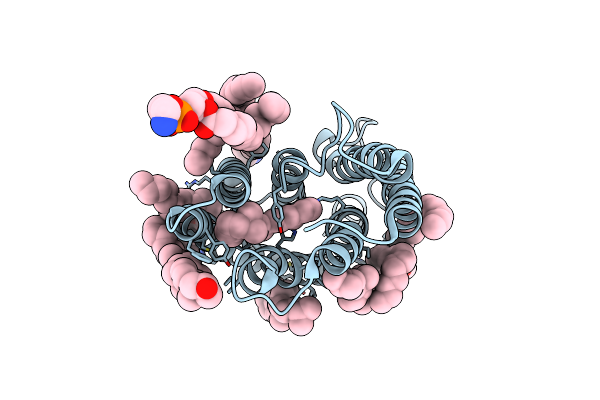 |
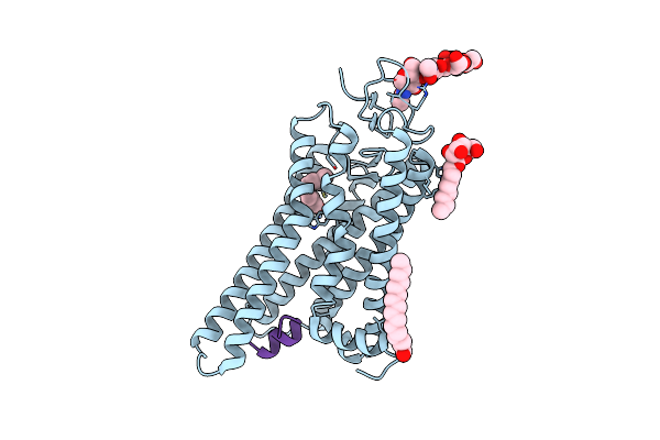
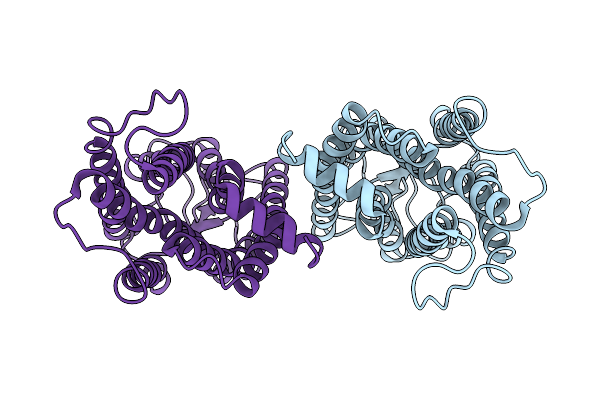
Kalium Channelrhodopsin 1 C110A Mutant From Hyphochytrium Catenoides, Dark State
Organism: Hyphochytrium catenoides
Method: ELECTRON MICROSCOPY Release Date: 2025-02-19 Classification: MEMBRANE PROTEIN Ligands: RET, CLR, PEE |
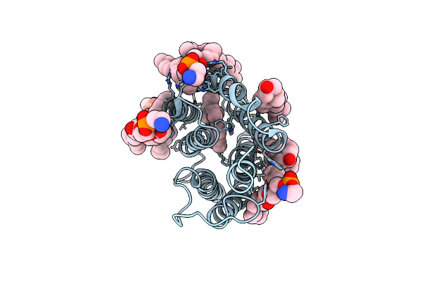 |
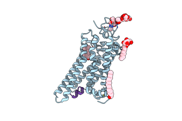
Kalium Channelrhodopsin 1 C110A Mutant From Hyphochytrium Catenoides, Laser-Flash-Illuminated
Organism: Hyphochytrium catenoides
Method: ELECTRON MICROSCOPY Release Date: 2025-02-19 Classification: MEMBRANE PROTEIN Ligands: RET, CLR, PEE |
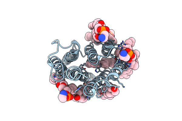 |
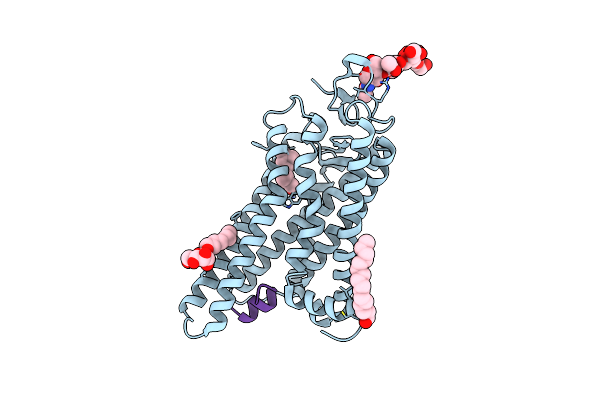
Kalium Channelrhodopsin 1 C110A Mutant From Hyphochytrium Catenoides, Continuous Illumination State
Organism: Hyphochytrium catenoides
Method: ELECTRON MICROSCOPY Release Date: 2025-02-19 Classification: MEMBRANE PROTEIN Ligands: RET, CLR, PEE |
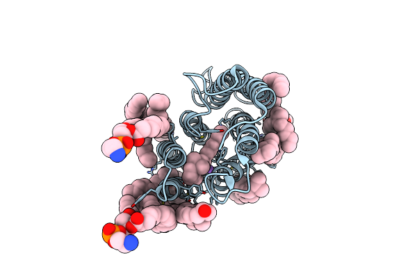 |
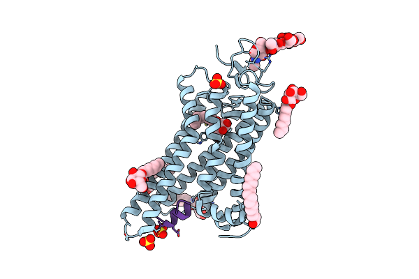
Kalium Channelrhodopsin 1 From Hyphochytrium Catenoides (Hckcr1) Embedded In Peptidisc
Organism: Hyphochytrium catenoides
Method: ELECTRON MICROSCOPY Release Date: 2023-07-26 Classification: TRANSPORT PROTEIN Ligands: RET, NA, CLR, PEE |
 |
Cation Channelrhodopsin From Hyphochytrium Catenoides (Hcccr) Embedded In Peptidisc
Organism: Hyphochytrium catenoides
Method: ELECTRON MICROSCOPY Release Date: 2023-07-26 Classification: TRANSPORT PROTEIN Ligands: RET, NA, CLR, PEE |
 |
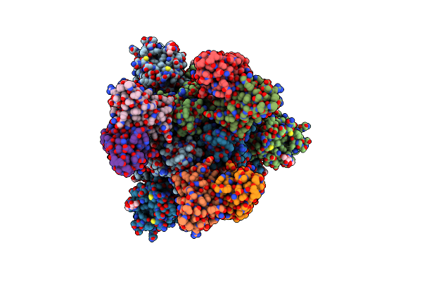
Organism: Homo sapiens, Severe acute respiratory syndrome coronavirus 2
Method: X-RAY DIFFRACTION Resolution:2.89 Å Release Date: 2023-07-19 Classification: ANTIVIRAL PROTEIN/IMMUNE SYSTEM Ligands: GOL, ZN, NAG |
 |
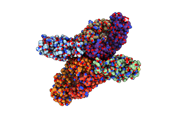
Sars-Cov-2 Spike Glycoprotein In Complex With The Ico-Hu23 Neutralizing Antibody Fab Fragment
Organism: Severe acute respiratory syndrome coronavirus 2, Homo sapiens
Method: ELECTRON MICROSCOPY Release Date: 2023-07-19 Classification: VIRAL PROTEIN/IMMUNE SYSTEM Ligands: NAG |
 |
Organism: Thermoplasmatales archaeon sg8-52-1
Method: X-RAY DIFFRACTION Resolution:1.97 Å Release Date: 2022-09-07 Classification: MEMBRANE PROTEIN Ligands: RET, CL, D12 |
 |
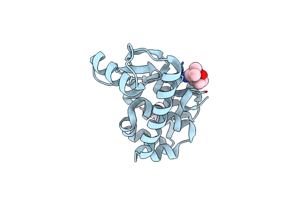
Organism: Rhodopseudomonas palustris
Method: X-RAY DIFFRACTION Resolution:1.75 Å Release Date: 2021-04-07 Classification: HYDROLASE Ligands: CL |
 |
Organism: Rhodopseudomonas palustris
Method: X-RAY DIFFRACTION Resolution:1.75 Å Release Date: 2021-04-07 Classification: HYDROLASE Ligands: CL |
 |
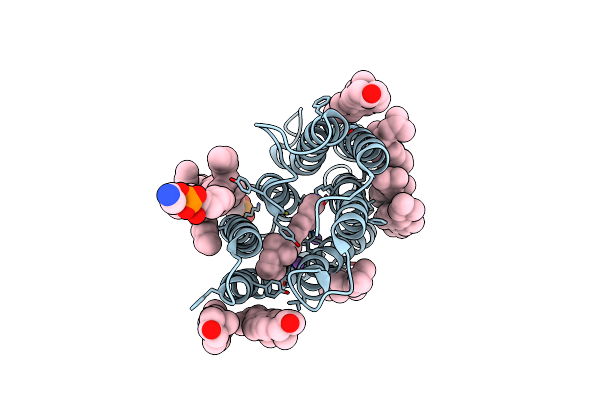
Co-Bound Sperm Whale Myoglobin Measured By Serial Synchrotron Crystallography
Organism: Physeter catodon
Method: X-RAY DIFFRACTION Resolution:1.75 Å Release Date: 2021-04-07 Classification: METAL BINDING PROTEIN Ligands: HEM, SO4, CMO |
 |
Co-Bound Sperm Whale Myoglobin Measured By Serial Femtosecond Crystallography
Organism: Physeter catodon
Method: X-RAY DIFFRACTION Resolution:1.75 Å Release Date: 2021-04-07 Classification: METAL BINDING PROTEIN Ligands: HEM, SO4, CMO |
 |
Organism: Enterobacteria phage t4
Method: X-RAY DIFFRACTION Resolution:1.50 Å Release Date: 2020-10-07 Classification: HYDROLASE Ligands: QPM |
 |
Organism: Mastigocladopsis repens
Method: X-RAY DIFFRACTION Resolution:2.50 Å Release Date: 2020-07-29 Classification: MEMBRANE PROTEIN Ligands: RET, BOG |
 |
Organism: Mastigocladopsis repens
Method: X-RAY DIFFRACTION Resolution:2.33 Å Release Date: 2020-07-29 Classification: MEMBRANE PROTEIN Ligands: RET, CL, C14, BOG, D10 |
 |
Organism: Bos taurus
Method: X-RAY DIFFRACTION Resolution:2.91 Å Release Date: 2020-07-01 Classification: SIGNALING PROTEIN Ligands: BOG, PLM, 64Z |
 |
Organism: Bos taurus
Method: X-RAY DIFFRACTION Resolution:2.90 Å Release Date: 2020-07-01 Classification: SIGNALING PROTEIN Ligands: BOG, PLM, NZZ |
 |
Organism: Bos taurus
Method: X-RAY DIFFRACTION Resolution:3.19 Å Release Date: 2020-06-24 Classification: SIGNALING PROTEIN Ligands: ODM, BOG, PLM |
 |
Organism: Bos taurus
Method: X-RAY DIFFRACTION Resolution:2.71 Å Release Date: 2020-02-12 Classification: SIGNALING PROTEIN Ligands: BOG, PLM, SO4 |
 |
Cryo-Em Structure Of The Native Rhodopsin Dimer From Rod Photoreceptor Cells
Organism: Bos taurus
Method: ELECTRON MICROSCOPY Release Date: 2019-08-21 Classification: SIGNALING PROTEIN |