
Search Count: 19
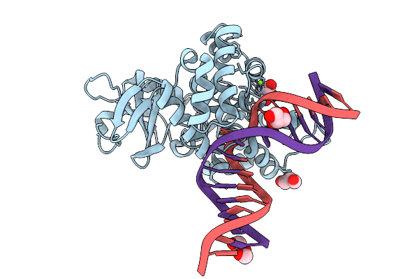 |
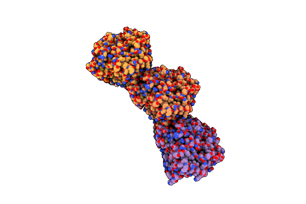
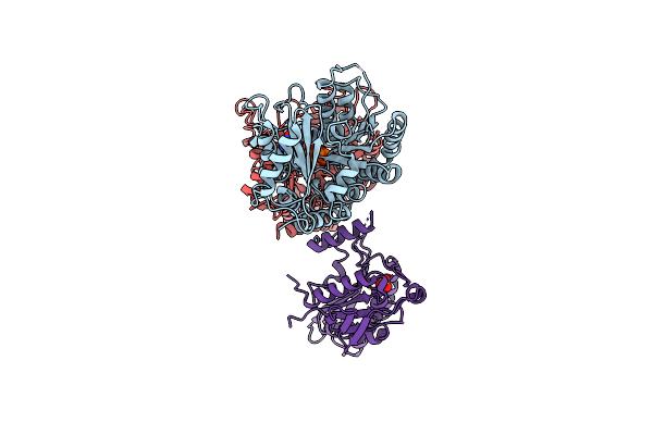
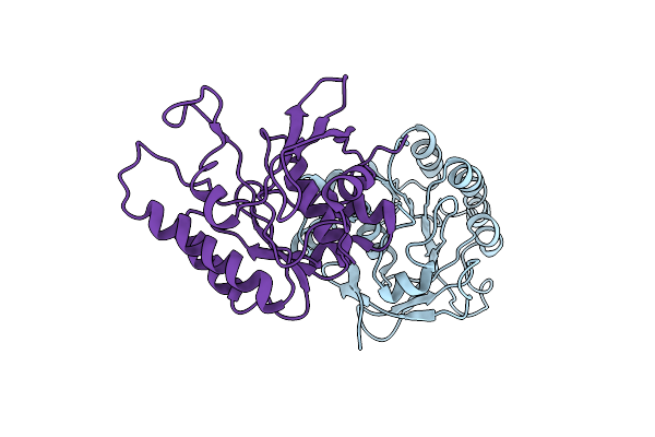
Crystal Structure Of Human 8-Oxoguanine Glycosylase K249H Mutant Bound To The Substrate 8-Oxoguanine Dna At Ph 8.0 Under 277 K
Organism: Homo sapiens
Method: X-RAY DIFFRACTION Release Date: 2025-07-23 Classification: DNA/HYDROLASE Ligands: PEG, GOL, MG, NA |
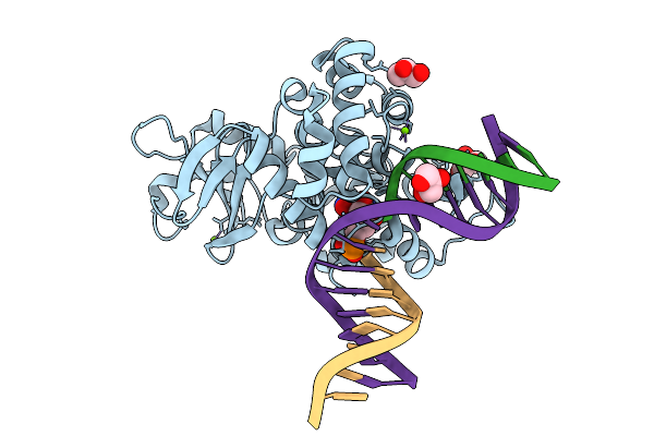 |
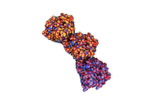
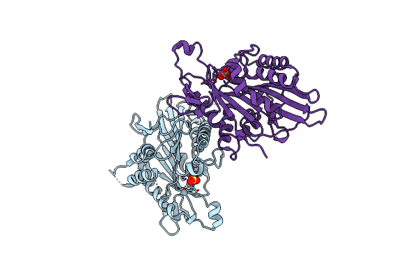
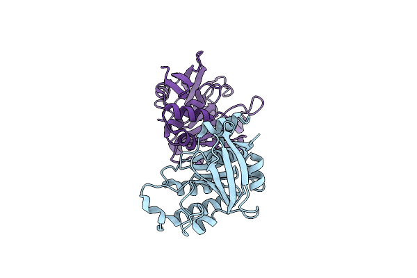
Crystal Structure Of Human 8-Oxoguanine Glycosylase K249H Mutant Bound To The Reaction Intermediate Derived From The Crystal Soaked Into The Solution At Ph 4.0 Under 277 K For 24 Hourss
Organism: Homo sapiens
Method: X-RAY DIFFRACTION Release Date: 2025-07-23 Classification: DNA/HYDROLASE Ligands: A1LXK, MG, GOL |
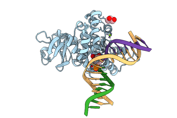 |
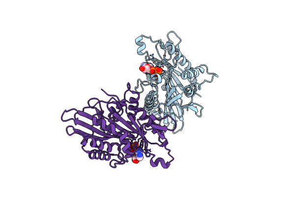
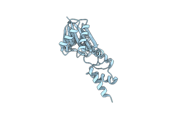
Crystal Structure Of Human 8-Oxoguanine Glycosylase K249H Mutant Bound To The Reaction Intermediate Derived From The Crystal Soaked Into The Solution At Ph 4.0 Under 277 K For 2.5 Hours
Organism: Homo sapiens
Method: X-RAY DIFFRACTION Release Date: 2025-07-23 Classification: DNA/HYDROLASE Ligands: A1LXK, MG, GOL |
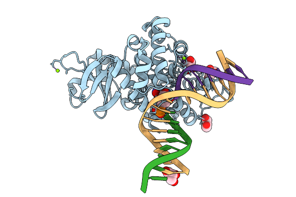 |
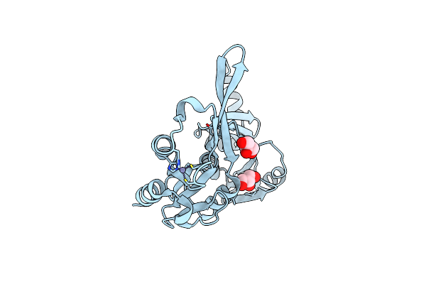
Crystal Structure Of Human 8-Oxoguanine Glycosylase K249H Mutant Bound To The Reaction Intermediate Derived From The Crystal Soaked Into The Solution At Ph 4.0 Under 298 K For 3 Weeks
Organism: Homo sapiens
Method: X-RAY DIFFRACTION Release Date: 2025-07-23 Classification: DNA/HYDROLASE Ligands: A1LXK, GOL, MG, NA |
 |
Organism: Mus musculus
Method: X-RAY DIFFRACTION Resolution:2.57 Å Release Date: 2021-12-29 Classification: MOTOR PROTEIN Ligands: ADP, AF3, MG |
 |
Organism: Mus musculus
Method: X-RAY DIFFRACTION Resolution:1.76 Å Release Date: 2021-12-29 Classification: MOTOR PROTEIN Ligands: AF3, MG, ADP |
 |
Organism: Sus scrofa
Method: ELECTRON MICROSCOPY Release Date: 2018-10-10 Classification: STRUCTURAL PROTEIN Ligands: GTP, MG, GDP |
 |
Organism: Sus scrofa
Method: ELECTRON MICROSCOPY Release Date: 2018-10-10 Classification: STRUCTURAL PROTEIN Ligands: GTP, MG, GDP |
 |
Organism: Sus scrofa
Method: ELECTRON MICROSCOPY Release Date: 2018-10-10 Classification: STRUCTURAL PROTEIN Ligands: GTP, MG, G2P |
 |
Kinesin-8 Motor, Kif19A, In The Nucleotide-Free State Complexed With Gdp-Taxol Microtubule
Organism: Mus musculus
Method: ELECTRON MICROSCOPY Release Date: 2016-09-28 Classification: MOTOR PROTEIN |
 |
Organism: Mus musculus
Method: X-RAY DIFFRACTION Resolution:2.72 Å Release Date: 2016-09-28 Classification: MOTOR PROTEIN Ligands: ADP, MG |
 |
Organism: Sus scrofa
Method: ELECTRON MICROSCOPY Release Date: 2016-09-28 Classification: STRUCTURAL PROTEIN Ligands: MG, GTP, GDP |
 |
Organism: Mus musculus, Sus scrofa
Method: ELECTRON MICROSCOPY Release Date: 2015-04-01 Classification: STRUCTURAL PROTEIN/MOTOR PROTEIN Ligands: MG, GTP, G2P, SO4 |
 |
Organism: Mus musculus
Method: X-RAY DIFFRACTION Resolution:2.86 Å Release Date: 2015-04-01 Classification: MOTOR PROTEIN Ligands: SO4 |
 |
Organism: Mus musculus
Method: X-RAY DIFFRACTION Resolution:2.70 Å Release Date: 2015-04-01 Classification: MOTOR PROTEIN Ligands: ADP |
 |
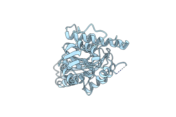
Crystal Structure Of Tbp-Interacting Protein (Tk-Tip26) And Implications For Its Inhibition Mechanism Of The Interaction Between Tbp And Tata-Dna
Organism: Thermococcus kodakarensis
Method: X-RAY DIFFRACTION Resolution:2.30 Å Release Date: 2006-02-14 Classification: TRANSCRIPTION REGULATOR Ligands: ZN, GOL |
 |
Organism: Thermococcus kodakarensis
Method: X-RAY DIFFRACTION Resolution:2.60 Å Release Date: 2005-08-09 Classification: DNA BINDING PROTEIN |
 |
Organism: Thermococcus kodakarensis
Method: X-RAY DIFFRACTION Resolution:2.20 Å Release Date: 2005-08-09 Classification: DNA BINDING PROTEIN |
 |
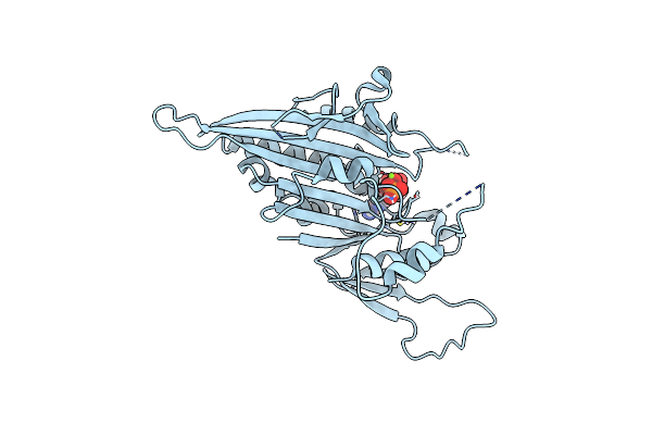
Crystal Structure Of Type 2 Ribonuclease H From Hyperthermophilic Archaeon, Thermococcus Kodakaraensis Kod1
Organism: Thermococcus kodakarensis
Method: X-RAY DIFFRACTION Resolution:2.00 Å Release Date: 2001-04-18 Classification: HYDROLASE |