
Search Count: 35
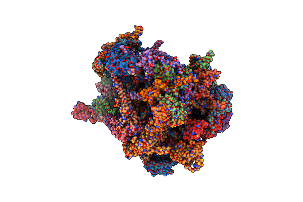 |
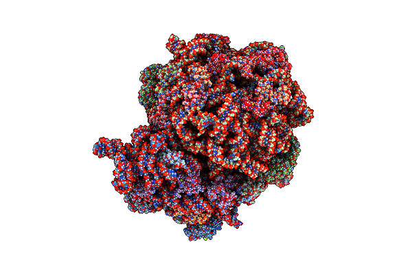
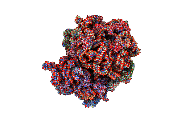
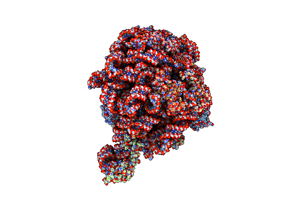
Organism: Homo sapiens
Method: ELECTRON MICROSCOPY Release Date: 2025-04-23 Classification: RIBOSOME/ANTIBIOTIC Ligands: ZN, MG, ZLD |
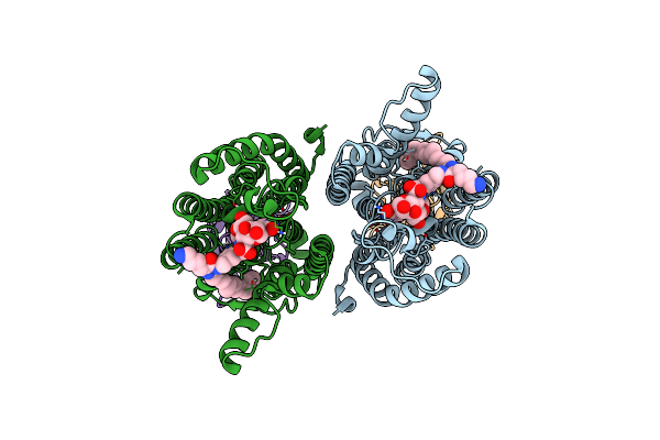 |
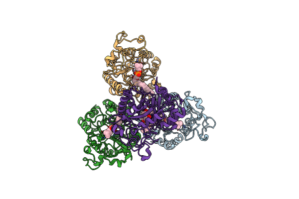
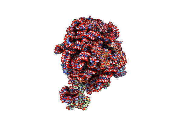
Organism: Aquifex aeolicus vf5, Lama glama
Method: ELECTRON MICROSCOPY Release Date: 2024-06-26 Classification: TRANSFERASE Ligands: A1AI1 |
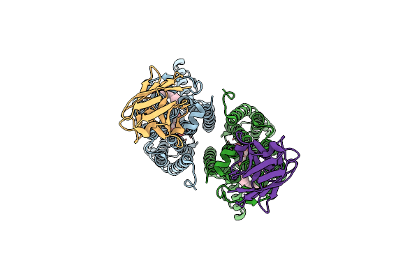 |
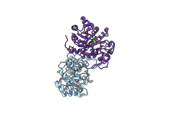
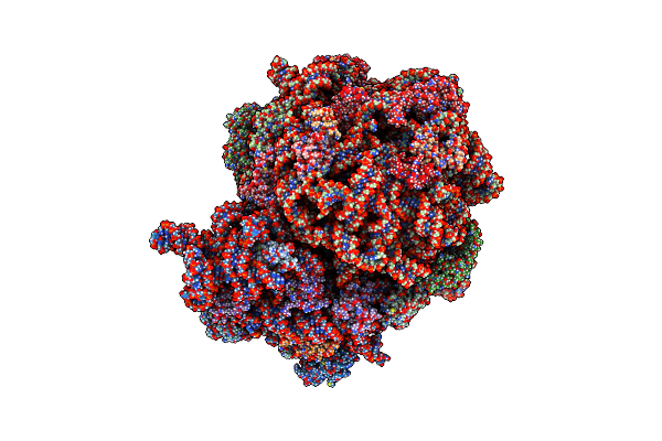
Organism: Lama glama, Aquifex aeolicus vf5
Method: ELECTRON MICROSCOPY Release Date: 2024-06-26 Classification: TRANSFERASE Ligands: A1AI2 |
 |
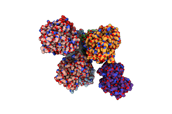
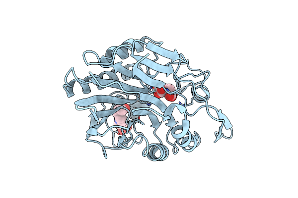
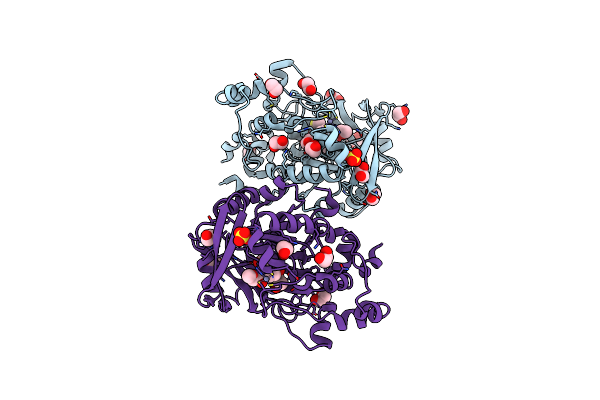
Structure Of Glycerophosphoethanolamine Ethanolaminephosphodiesterase From Streptomyces Sanglieri
Organism: Streptomyces sanglieri
Method: X-RAY DIFFRACTION Resolution:2.10 Å Release Date: 2024-04-03 Classification: HYDROLASE Ligands: CA, GOL |
 |
Organism: Thermocrispum sp. rd004668
Method: X-RAY DIFFRACTION Resolution:2.57 Å Release Date: 2023-02-08 Classification: HYDROLASE |
 |
Organism: Thermocrispum sp. rd004668
Method: X-RAY DIFFRACTION Resolution:2.29 Å Release Date: 2023-02-08 Classification: HYDROLASE |
 |
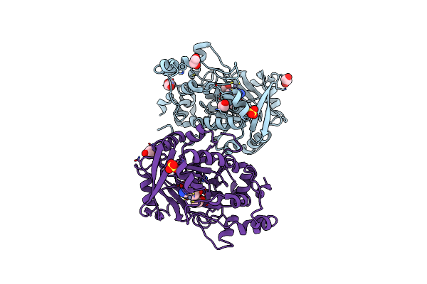
Complex Structure Of Lysoplasmalogen Specific Phopholipase D, F211L Mutant With Lpc
Organism: Thermocrispum sp. rd004668
Method: X-RAY DIFFRACTION Resolution:2.69 Å Release Date: 2023-02-08 Classification: HYDROLASE Ligands: KIP |
 |
Organism: Thermocrispum sp. rd004668
Method: X-RAY DIFFRACTION Resolution:2.91 Å Release Date: 2023-01-04 Classification: HYDROLASE Ligands: CA |
 |
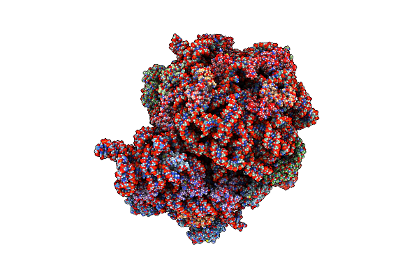
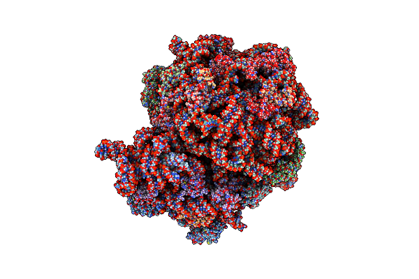
Organism: Escherichia coli
Method: ELECTRON MICROSCOPY Release Date: 2022-03-02 Classification: RIBOSOME Ligands: MG, RD8, ZN |
 |
Organism: Escherichia coli
Method: ELECTRON MICROSCOPY Release Date: 2022-03-02 Classification: RIBOSOME Ligands: MG, RD8, ZN |
 |
Organism: Escherichia coli
Method: ELECTRON MICROSCOPY Release Date: 2022-01-19 Classification: RIBOSOME Ligands: MG, RD8, ZN |
 |
Organism: Escherichia coli
Method: ELECTRON MICROSCOPY Release Date: 2021-12-15 Classification: RIBOSOME Ligands: MG, NA, ZN |
 |
Organism: Escherichia coli
Method: ELECTRON MICROSCOPY Release Date: 2021-11-17 Classification: RIBOSOME/ANTIBIOTIC Ligands: MG, ZLD, ZN |
 |
Organism: Escherichia coli
Method: ELECTRON MICROSCOPY Release Date: 2021-11-17 Classification: RIBOSOME/ANTIBIOTIC Ligands: MG, ZLD, ZN |
 |
Organism: Homo sapiens
Method: SOLUTION NMR Release Date: 2021-03-03 Classification: GENE REGULATION Ligands: ZN |
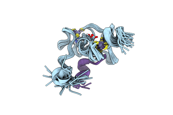 |
Solution Structure Of The Phd1 Domain Of Histone Demethylase Kdm5A In Complex With A Histone H3(1-10) Peptide
Organism: Homo sapiens
Method: SOLUTION NMR Release Date: 2021-03-03 Classification: GENE REGULATION Ligands: ZN |
 |
Organism: Streptomyces griseocarneus
Method: X-RAY DIFFRACTION Resolution:2.00 Å Release Date: 2020-10-28 Classification: HYDROLASE Ligands: EPE, SIN |
 |
Organism: Escherichia coli, Escherichia coli
Method: ELECTRON MICROSCOPY Release Date: 2020-01-22 Classification: RIBOSOME Ligands: MG, NA, ZN |
 |
Organism: Homo sapiens
Method: X-RAY DIFFRACTION Resolution:2.39 Å Release Date: 2016-01-13 Classification: OXIDOREDUCTASE Ligands: MN, ZN, EDO, SO4, VAO |
 |
Organism: Homo sapiens
Method: X-RAY DIFFRACTION Resolution:2.15 Å Release Date: 2016-01-13 Classification: OXIDOREDUCTASE Ligands: SO4, 7WH, EDO, ZN, MN, DMS |