
Search Count: 15
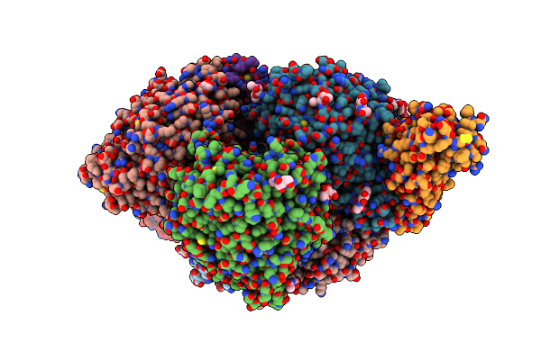 |
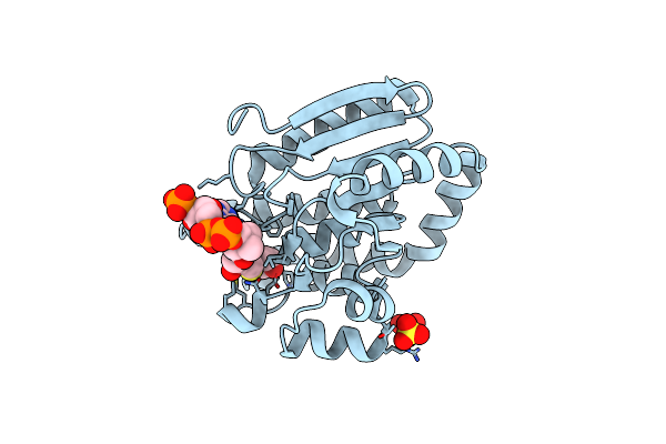
Crystal Structure Of Affinity-Enhancing And Catalytically Inactive Ace2 In Complex With Sars-Cov-2 Rbd
Organism: Homo sapiens, Severe acute respiratory syndrome coronavirus 2
Method: X-RAY DIFFRACTION Resolution:3.54 Å Release Date: 2021-12-15 Classification: Hydrolase/Viral protein Ligands: NAG, ZN |
 |
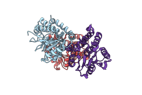 |
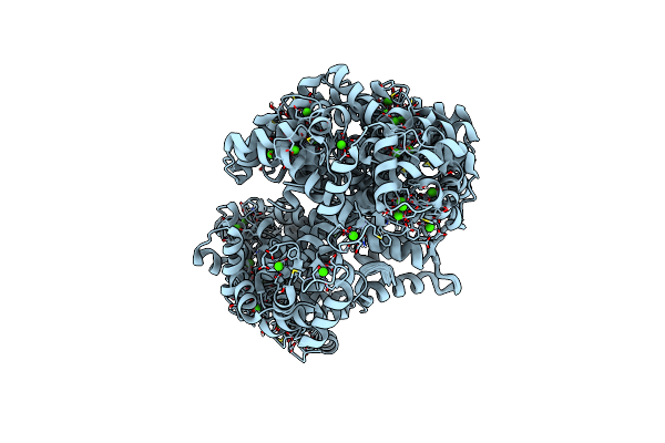
Organism: Mycobacterium tuberculosis
Method: X-RAY DIFFRACTION Resolution:1.25 Å Release Date: 2013-05-29 Classification: ISOMERASE Ligands: CL |
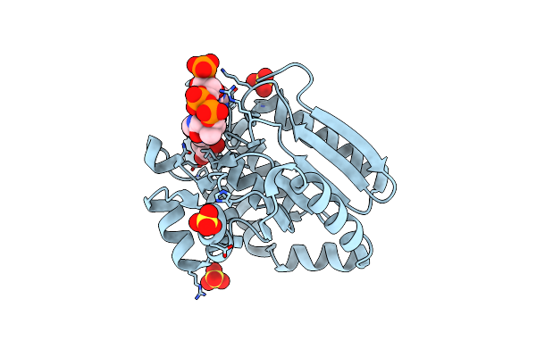 |
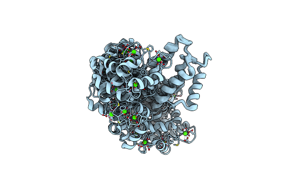
Crystal Structure Of The Mtb Enoyl Coa Isomerase (Rv0632C)In Complex With Acetoacetyl Coa
Organism: Mycobacterium tuberculosis
Method: X-RAY DIFFRACTION Resolution:1.83 Å Release Date: 2013-05-29 Classification: ISOMERASE Ligands: CAA, SO4 |
 |
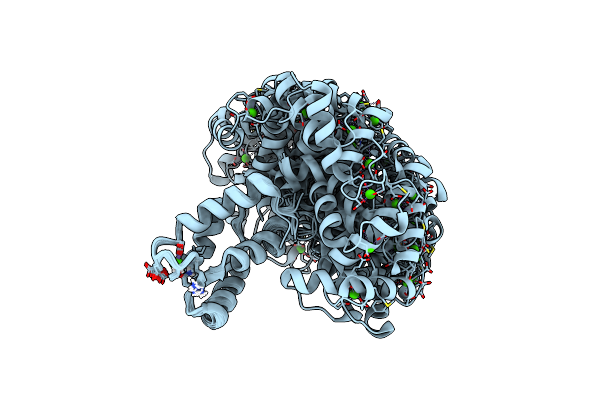
Crystal Structure Of The Mtb Enoyl Coa Isomerase (Rv0632C) In Complex With Hydroxybutyrl Coa
Organism: Mycobacterium tuberculosis
Method: X-RAY DIFFRACTION Resolution:1.80 Å Release Date: 2013-05-29 Classification: ISOMERASE Ligands: SO4, 3HC |
 |
Crystal Structure Of The Mtb Enoyl Coa Isomerase In Complex With Hydroxyhexanoyl Coa
Organism: Mycobacterium tuberculosis
Method: X-RAY DIFFRACTION Resolution:1.85 Å Release Date: 2013-05-29 Classification: ISOMERASE Ligands: 3H9, SO4 |
 |
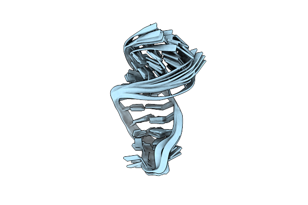
Organism: Drosophila melanogaster
Method: SOLUTION NMR Release Date: 2012-08-22 Classification: METAL BINDING PROTEIN Ligands: CA |
 |
Organism: Drosophila melanogaster
Method: SOLUTION NMR Release Date: 2012-08-22 Classification: METAL BINDING PROTEIN Ligands: CA |
 |
Organism: Drosophila melanogaster
Method: SOLUTION NMR Release Date: 2012-08-22 Classification: METAL BINDING PROTEIN Ligands: CA |
 |
Crystal Structure Of Wlac Mutant Of Dimerisation Domain Of Nf-Kb P50 Transcription Factor
Organism: Mus musculus
Method: X-RAY DIFFRACTION Resolution:1.89 Å Release Date: 2004-08-17 Classification: TRANSCRIPTION |
 |
Crystal Structure Of Mlav Mutant Of Dimerisation Domain Of Nf-Kb P50 Transcription Factor
Organism: Mus musculus
Method: X-RAY DIFFRACTION Resolution:1.90 Å Release Date: 2004-08-17 Classification: TRANSCRIPTION |
 |
Crystal Structure Of Ilac Mutant Of Dimerisation Domain Of Nf-Kb P50 Transcription Factor
Organism: Mus musculus
Method: X-RAY DIFFRACTION Resolution:1.90 Å Release Date: 2004-08-17 Classification: TRANSCRIPTION |
 |
Crystal Structure Of Mlac Mutant Of Dimerisation Domain Of Nf-Kb P50 Transcription Factor
Organism: Mus musculus
Method: X-RAY DIFFRACTION Resolution:1.90 Å Release Date: 2004-08-17 Classification: TRANSCRIPTION |
 |
Crystal Structure Of Ylgv Mutant Of Dimerisation Domain Of Nf-Kb P50 Transcription Factor
Organism: Mus musculus
Method: X-RAY DIFFRACTION Resolution:2.20 Å Release Date: 2004-08-17 Classification: TRANSCRIPTION |
 |
Crystal Structure Of Mlam Mutant Of Dimerisation Domain Of Nf-Kb P50 Transcription Factor
Organism: Mus musculus
Method: X-RAY DIFFRACTION Resolution:2.70 Å Release Date: 2004-08-17 Classification: TRANSCRIPTION |