
Search Count: 18
 |
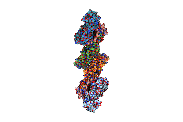
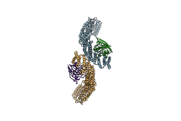
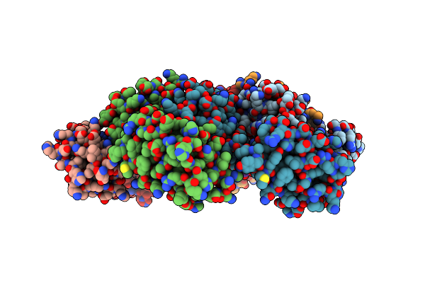
Cryo-Em Structure Of The Tripartite Atp-Independent Periplasmic (Trap) Transporter Siaqm From Photobacterium Profundum In A Nanodisc
Organism: Photobacterium profundum ss9, Helicobacter pylori, Synthetic construct
Method: ELECTRON MICROSCOPY Release Date: 2023-03-15 Classification: TRANSPORT PROTEIN Ligands: OCT, PTY, TWT, D10, NA, TRD, HEX |
 |
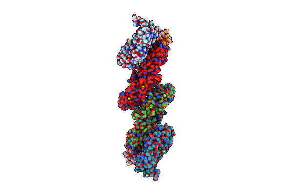
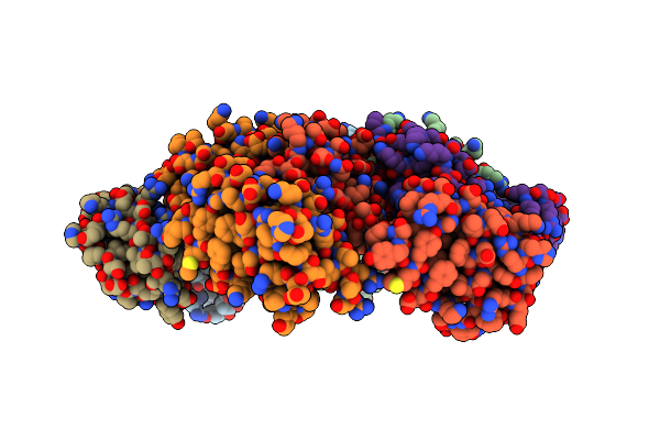
Cryo-Em Structure Of The Tripartite Atp-Independent Periplasmic (Trap) Transporter Siaqm From Photobacterium Profundum In Amphipol
Organism: Photobacterium profundum ss9, Helicobacter pylori, Synthetic construct
Method: ELECTRON MICROSCOPY Release Date: 2022-12-21 Classification: TRANSPORT PROTEIN |
 |
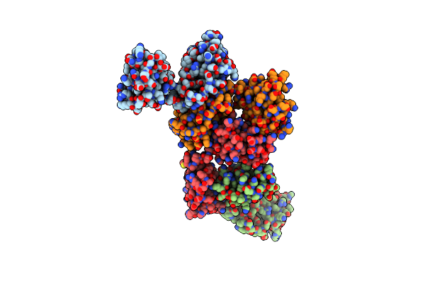
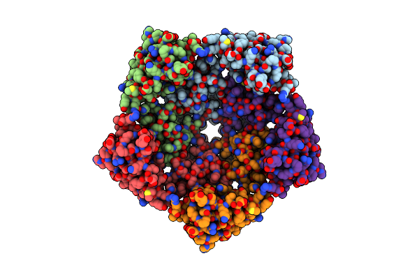
Structure Of The Sialic Acid Bound Tripartite Atp-Independent Periplasmic (Trap) Periplasmic Component Siap From Photobacterium Profundum
Organism: Photobacterium profundum
Method: X-RAY DIFFRACTION Resolution:1.04 Å Release Date: 2022-12-14 Classification: TRANSPORT PROTEIN Ligands: SLB, SO4 |
 |
Organism: Homo sapiens
Method: ELECTRON MICROSCOPY Resolution:9.80 Å Release Date: 2021-03-03 Classification: ENDOCYTOSIS Ligands: GTP, MG |
 |
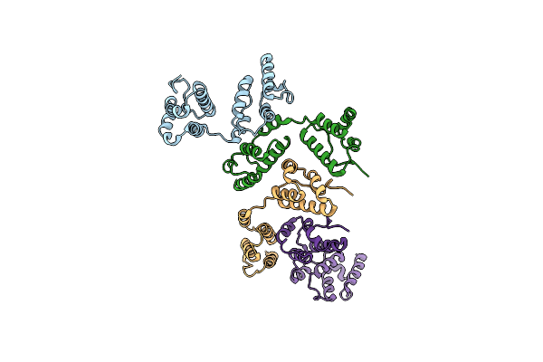
Vps26 Dimer Region Of Metazoan Membrane-Assembled Retromer:Snx3 Complex Modelled With Human Proteins
Organism: Homo sapiens, Mus musculus
Method: ELECTRON MICROSCOPY Release Date: 2021-03-03 Classification: ENDOCYTOSIS Ligands: PIB |
 |
Vps26 Dimer Region Of The Fungal Membrane-Assembled Retromer:Grd19 Complex.
Organism: Chaetomium thermophilum (strain dsm 1495 / cbs 144.50 / imi 039719), Chaetomium thermophilum var. thermophilum dsm 1495
Method: ELECTRON MICROSCOPY Release Date: 2021-03-03 Classification: ENDOCYTOSIS Ligands: PIB |
 |
Vps35/Vps29 Arch Of Metazoan Membrane-Assembled Retromer:Snx3 Complex Modelled With Human Proteins
Organism: Homo sapiens
Method: ELECTRON MICROSCOPY Release Date: 2021-02-10 Classification: ENDOCYTOSIS |
 |
Organism: Chaetomium thermophilum (strain dsm 1495 / cbs 144.50 / imi 039719)
Method: ELECTRON MICROSCOPY Release Date: 2021-02-10 Classification: ENDOCYTOSIS |
 |
Vps35/Vps29 Arch Of Fungal Membrane-Assembled Retromer:Vps5 (Snx-Bar) Complex.
Organism: Chaetomium thermophilum (strain dsm 1495 / cbs 144.50 / imi 039719)
Method: ELECTRON MICROSCOPY Release Date: 2021-02-10 Classification: ENDOCYTOSIS |
 |
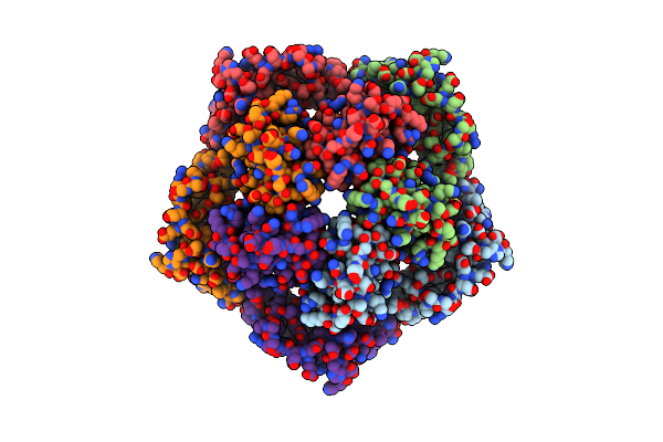
A Model Of The Eiav Ca-Sp Hexamer (C2) From Gag-Deltama Tubes Assembled At Ph8
Organism: Equine infectious anemia virus
Method: ELECTRON MICROSCOPY Release Date: 2020-01-15 Classification: VIRAL PROTEIN |
 |
A Model Of The Eiav Ca-Sp Hexamer (C2) From Gag-Deltama Tubes Assembled At Ph6
Organism: Equine infectious anemia virus
Method: ELECTRON MICROSCOPY Release Date: 2020-01-15 Classification: VIRAL PROTEIN |
 |
A Model Of The Eiav Ca-Sp Hexamer (C6) From Gag-Deltama Spheres Assembled At Ph6
Organism: Equine infectious anemia virus
Method: ELECTRON MICROSCOPY Release Date: 2020-01-15 Classification: VIRAL PROTEIN |
 |
Organism: Drosophila melanogaster
Method: ELECTRON MICROSCOPY Release Date: 2020-01-01 Classification: VIRUS LIKE PARTICLE Ligands: ZN |
 |
Organism: Drosophila melanogaster
Method: ELECTRON MICROSCOPY Release Date: 2020-01-01 Classification: VIRUS LIKE PARTICLE |
 |
Organism: Drosophila melanogaster
Method: ELECTRON MICROSCOPY Release Date: 2020-01-01 Classification: VIRUS LIKE PARTICLE |
 |
Organism: Drosophila melanogaster
Method: ELECTRON MICROSCOPY Release Date: 2020-01-01 Classification: VIRUS LIKE PARTICLE Ligands: ZN |
 |
Organism: Drosophila melanogaster
Method: ELECTRON MICROSCOPY Release Date: 2020-01-01 Classification: VIRUS LIKE PARTICLE |
 |
Organism: Drosophila melanogaster
Method: ELECTRON MICROSCOPY Release Date: 2020-01-01 Classification: VIRUS LIKE PARTICLE |