
Search Count: 94
 |
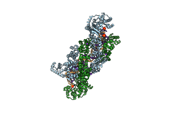
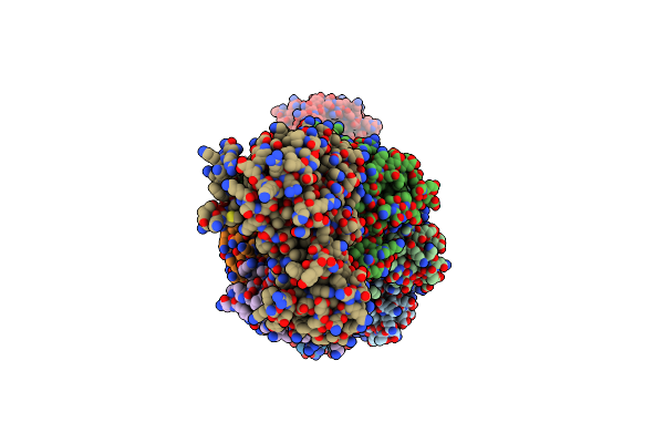
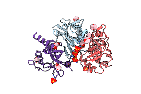
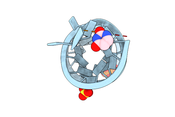
Structure Of M. Kandleri Topoisomerase V In Complex With Dna. 38 Base Pair Symmetric Dna Complex
Organism: Methanopyrus kandleri, Synthetic construct
Method: X-RAY DIFFRACTION Resolution:3.52 Å Release Date: 2022-08-31 Classification: DNA BINDING PROTEIN/DNA Ligands: K |
 |
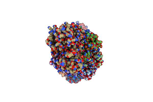
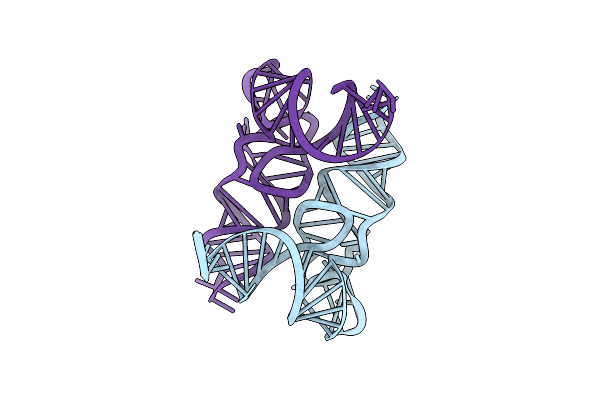
Structure Of M. Kandleri Topoisomerase V In Complex With Dna. 40 Base Pair Symmetric Dna Complex
Organism: Methanopyrus kandleri, Synthetic construct
Method: X-RAY DIFFRACTION Resolution:2.92 Å Release Date: 2022-08-31 Classification: DNA BINDING PROTEIN/DNA Ligands: K, PO4, PO3 |
 |
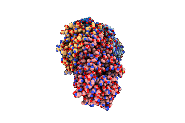
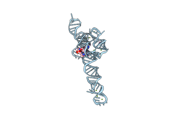
Structure Of M. Kandleri Topoisomerase V In Complex With Dna. 38 Base Pair Asymmetric Dna Complex
Organism: Methanopyrus kandleri, Synthetic construct
Method: X-RAY DIFFRACTION Resolution:3.24 Å Release Date: 2022-08-31 Classification: DNA BINDING PROTEIN/DNA Ligands: MG, K |
 |
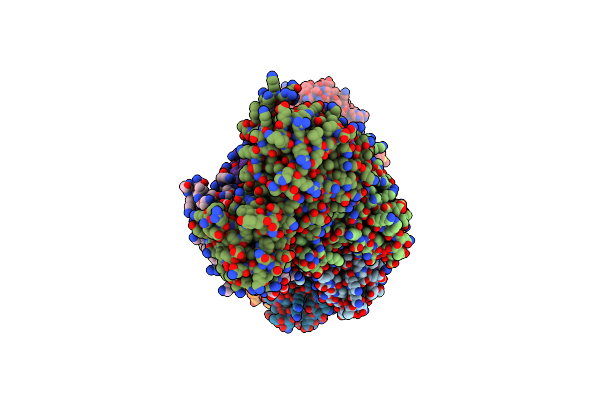
Structure Of M. Kandleri Topoisomerase V In Complex With Dna. 39 Base Pair Symmetric Dna Complex
Organism: Methanopyrus kandleri, Synthetic construct
Method: X-RAY DIFFRACTION Resolution:3.17 Å Release Date: 2022-08-31 Classification: DNA BINDING PROTEIN/DNA Ligands: K |
 |
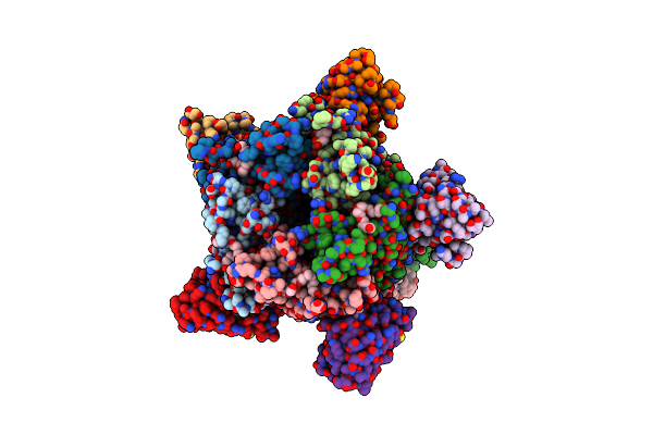
Organism: Staphylococcus epidermidis rp62a
Method: ELECTRON MICROSCOPY Release Date: 2022-07-06 Classification: RNA BINDING PROTEIN/RNA |
 |
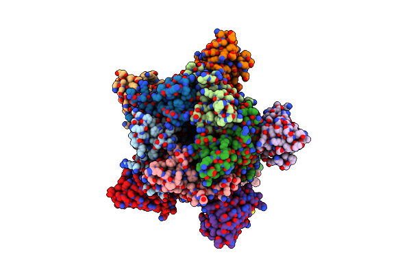
Staphylococcus Epidermidis Rp62A Crispr Effector Subcomplex With Non-Self Target Rna Bound
Organism: Staphylococcus epidermidis rp62a
Method: ELECTRON MICROSCOPY Release Date: 2022-07-06 Classification: HYDROLASE/RNA |
 |
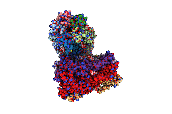
Staphylococcus Epidermidis Rp62A Crispr Effector Complex With Non-Self Target Rna 2
Organism: Staphylococcus epidermidis rp62a
Method: ELECTRON MICROSCOPY Release Date: 2022-07-06 Classification: HYDROLASE/RNA |
 |
Organism: Staphylococcus epidermidis rp62a
Method: ELECTRON MICROSCOPY Release Date: 2022-07-06 Classification: HYDROLASE/RNA |
 |
Staphylococcus Epidermidis Rp62A Crispr Tall Effector Complex With Bound Atp
Organism: Staphylococcus epidermidis rp62a
Method: ELECTRON MICROSCOPY Release Date: 2022-07-06 Classification: HYDROLASE/RNA Ligands: ATP |
 |
Staphylococcus Epidermidis Rp62A Crispr Short Effector Complex With Self Rna Target And Atp
Organism: Staphylococcus epidermidis, Staphylococcus epidermidis rp62a
Method: ELECTRON MICROSCOPY Release Date: 2022-07-06 Classification: HYDROLASE/RNA Ligands: ATP |
 |
Organism: Staphylococcus epidermidis rp62a
Method: ELECTRON MICROSCOPY Release Date: 2022-07-06 Classification: HYDROLASE/RNA |
 |
Organism: Thermotoga maritima (strain atcc 43589 / msb8 / dsm 3109 / jcm 10099)
Method: X-RAY DIFFRACTION Resolution:2.10 Å Release Date: 2022-02-09 Classification: RNA BINDING PROTEIN Ligands: DIO, SO4, GOL |
 |
Organism: Bacillus subtilis
Method: X-RAY DIFFRACTION Resolution:3.25 Å Release Date: 2020-06-10 Classification: RNA Ligands: MG, NCO, B1Z |
 |
Organism: Escherichia coli
Method: X-RAY DIFFRACTION Resolution:1.95 Å Release Date: 2020-01-01 Classification: RNA |
 |
Crystal Structure Of A Fragment Of E. Coli Trna(Asp) Consisting Of Its Acceptor Stem/T Stem-Loop. Long Unit Cell.
Organism: Escherichia coli
Method: X-RAY DIFFRACTION Resolution:1.75 Å Release Date: 2020-01-01 Classification: RNA Ligands: SO4 |
 |
Crystal Structure Of A Fragment Of E. Coli Trna(Asp) Consisting Of Its Acceptor Stem/T Stem-Loop. Short Unit Cell.
Organism: Escherichia coli
Method: X-RAY DIFFRACTION Resolution:1.60 Å Release Date: 2020-01-01 Classification: RNA Ligands: SO4, URA, DPO |
 |
X-Ray Structure Of A Pentameric Ligand Gated Ion Channel From Erwinia Chrysanthemi (Elic) 7'C Pore Mutant (L238C) In Complex With Nanobody 72
Organism: Dickeya chrysanthemi, Lama glama
Method: X-RAY DIFFRACTION Resolution:2.50 Å Release Date: 2019-10-09 Classification: MEMBRANE PROTEIN Ligands: P6G, PTY, LMT, MES, NA |
 |
X-Ray Structure Of A Pentameric Ligand Gated Ion Channel From Erwinia Chrysanthemi (Elic) Delta8 Truncation Mutant In Complex With Nanobody 72
Organism: Dickeya chrysanthemi, Lama glama
Method: X-RAY DIFFRACTION Resolution:2.78 Å Release Date: 2019-10-09 Classification: MEMBRANE PROTEIN |
 |
X-Ray Structure Of A Pentameric Ligand Gated Ion Channel From Erwinia Chrysanthemi (Elic) F16'S Pore Mutant (F247S) With Alternate M4 Conformation.
Organism: Dickeya chrysanthemi
Method: X-RAY DIFFRACTION Resolution:3.45 Å Release Date: 2019-10-09 Classification: MEMBRANE PROTEIN Ligands: LMT |
 |
Organism: Staphylococcus epidermidis (strain atcc 35984 / rp62a)
Method: X-RAY DIFFRACTION Resolution:2.40 Å Release Date: 2019-02-13 Classification: RNA BINDING PROTEIN Ligands: CA, SM |