
Search Count: 16
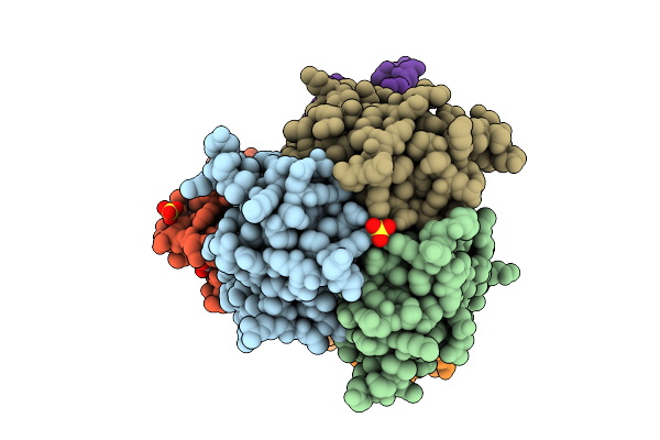 |
Organism: Homo sapiens, Squalus acanthias
Method: X-RAY DIFFRACTION Release Date: 2025-11-05 Classification: Cytokine/Immune System Ligands: EDO, SO4, NI |
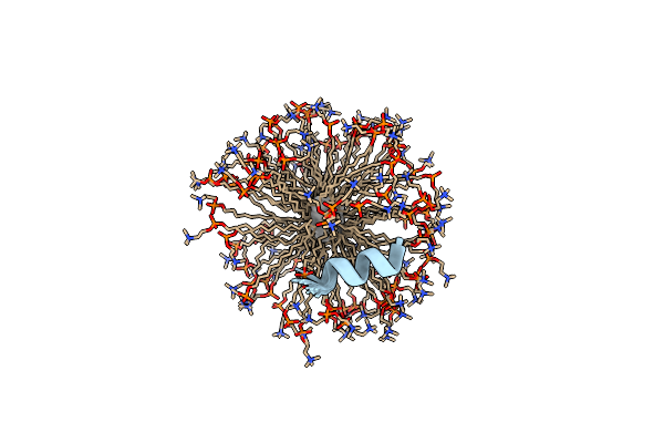 |
Organism: Anoplius samariensis
Method: SOLUTION NMR Release Date: 2014-12-03 Classification: ANTIMICROBIAL PROTEIN Ligands: UNX, DPV |
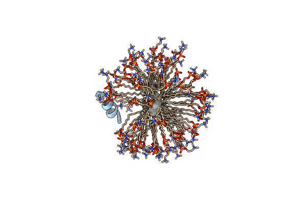 |
Organism: Anoplius samariensis
Method: SOLUTION NMR Release Date: 2014-12-03 Classification: ANTIMICROBIAL PROTEIN Ligands: UNX, DPV |
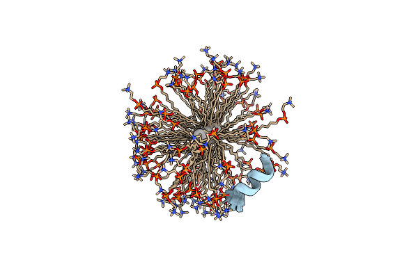 |
Organism: Anoplius samariensis
Method: SOLUTION NMR Release Date: 2014-12-03 Classification: ANTIMICROBIAL PROTEIN Ligands: UNX, DPV |
 |
Organism: Anoplius samariensis
Method: SOLUTION NMR Release Date: 2014-12-03 Classification: ANTIMICROBIAL PROTEIN Ligands: UNX, DPV |
 |
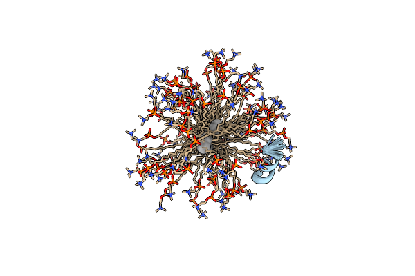
Plectasin:A Peptide Antibiotic With Therapeutic Potential From A Saprophytic Fungus
Organism: Pseudoplectania nigrella
Method: SOLUTION NMR Release Date: 2005-10-18 Classification: ANTIMICROBIAL PROTEIN |
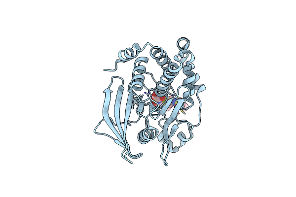 |
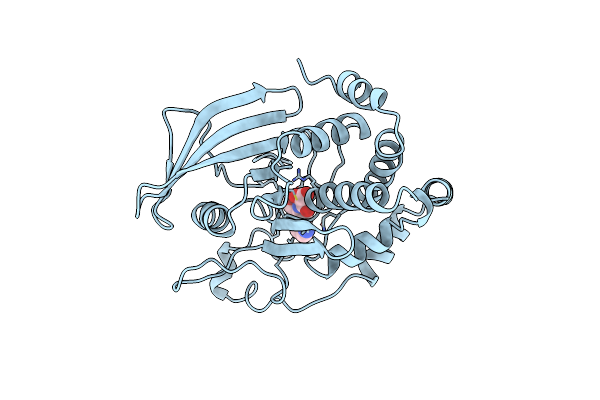
Crystal Structure Of Ptp1B Complexed With 7-(1,1-Dioxo-1H-Benzo[D]Isothiazol-3-Yloxymethyl)-2-(Oxalyl-Amino)-4,7-Dihydro-5H-Thieno[2,3-C]Pyran-3-Carboxylic Acid
Organism: Homo sapiens
Method: X-RAY DIFFRACTION Resolution:2.50 Å Release Date: 2002-05-08 Classification: HYDROLASE Ligands: DBD |
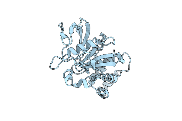 |
Organism: Homo sapiens
Method: X-RAY DIFFRACTION Resolution:2.56 Å Release Date: 2002-05-08 Classification: HYDROLASE |
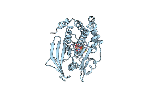 |
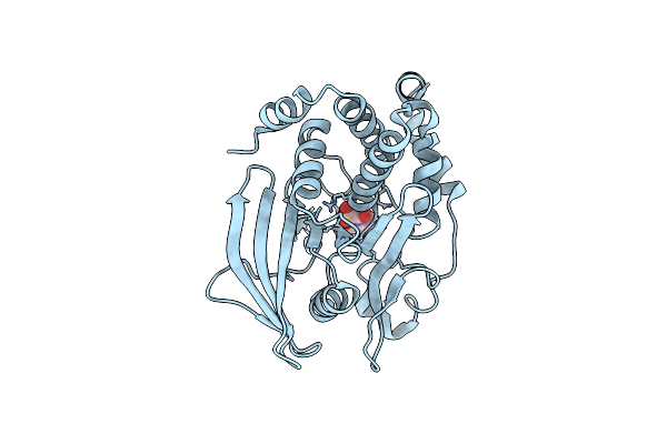
Residue 259 Is A Key Determinant Of Substrate Specificity Of Protein-Tyrosine Phosphatase 1B And Alpha
Organism: Homo sapiens
Method: X-RAY DIFFRACTION Resolution:2.13 Å Release Date: 2000-07-04 Classification: HYDROLASE Ligands: COL |
 |
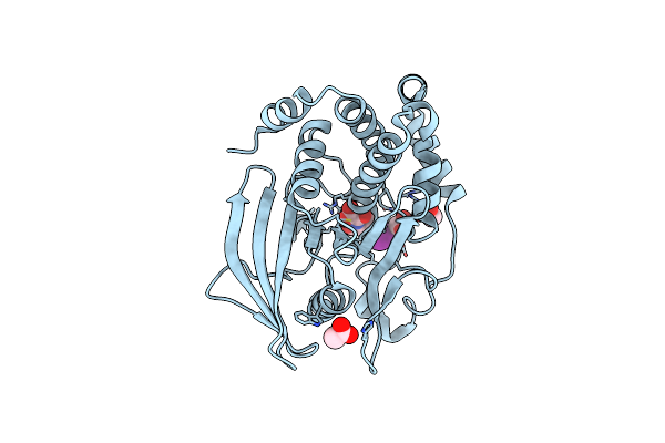
Crystal Structure Of Protein Tyrosine Phosphatase 1B Complexed With 6-(Oxalyl-Amino)-1H-Indole-5-Carboxylic Acid
Organism: Homo sapiens
Method: X-RAY DIFFRACTION Resolution:1.80 Å Release Date: 2000-05-03 Classification: HYDROLASE Ligands: OAI |
 |
Crystal Structure Of Protein Tyrosine Phosphatase 1B Complexed With 3-(Oxalyl-Amino)-Naphthalene-2-Carboxlic Acid
Organism: Homo sapiens
Method: X-RAY DIFFRACTION Resolution:2.35 Å Release Date: 2000-05-03 Classification: HYDROLASE Ligands: 761 |
 |
Crystal Structure Of Protein Tyrosine Phosphatase 1B Complexed With 2-(Oxalyl-Amino)-Benzoic Acid
Organism: Homo sapiens
Method: X-RAY DIFFRACTION Resolution:2.72 Å Release Date: 2000-05-03 Classification: HYDROLASE Ligands: OBA |
 |
Crystal Structure Of Protein Tyrosine Phosphatase 1B (R47V,D48N) Complexed With 2-(Oxalyl-Amino-4,7-Dihydro-5H-Thieno[2,3-C]Pyran-3-Carboxylic Acid
Organism: Homo sapiens
Method: X-RAY DIFFRACTION Resolution:2.30 Å Release Date: 2000-05-03 Classification: HYDROLASE Ligands: OPA |
 |
Crystal Structure Of Protein Tyrosine Phosphatase 1B Complexed With 2-(Oxalyl-Amino-4,7-Dihydro-5H-Thieno[2,3-C]Pyran-3-Carboxylic Acid
Organism: Homo sapiens
Method: X-RAY DIFFRACTION Resolution:2.10 Å Release Date: 2000-05-03 Classification: HYDROLASE Ligands: OPA |
 |
Crystal Structure Of Protein Tyrosine Phosphatase 1B Complexed With 2-(Oxalyl-Amino)-4,5,6,7-Tetrahydro-Thieno[2,3-C]Pyridine-3-Carboxylic Acid
Organism: Homo sapiens
Method: X-RAY DIFFRACTION Resolution:1.80 Å Release Date: 2000-05-03 Classification: HYDROLASE Ligands: OTA |
 |
Crystal Structure Of Protein Tyrosine Phosphatase 1B Complexed With 5-Iodo-2-(Oxalyl-Amino)-Benzoic Acid
Organism: Homo sapiens
Method: X-RAY DIFFRACTION Resolution:1.95 Å Release Date: 2000-03-15 Classification: HYDROLASE Ligands: ACT, 878 |