
Search Count: 22
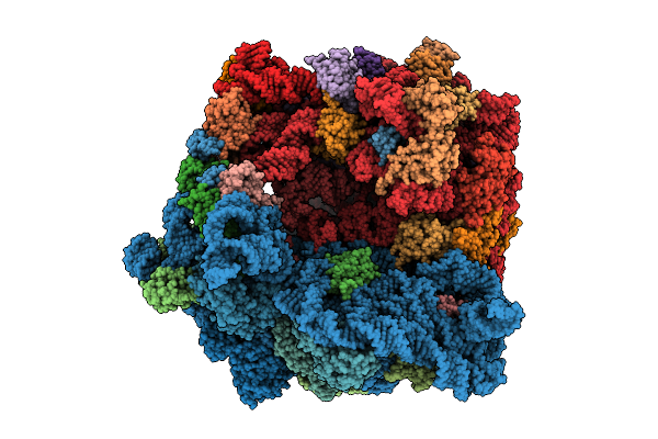 |
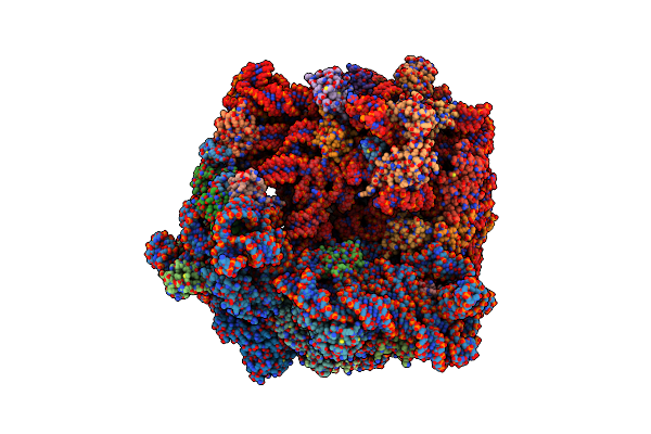
Structure Of The Bacterial Ribosome Without Hypoxia-Induced Rrna Modifications
Organism: Escherichia phage t4, Escherichia coli bw25113
Method: ELECTRON MICROSCOPY Release Date: 2025-11-12 Classification: RIBOSOME Ligands: MG |
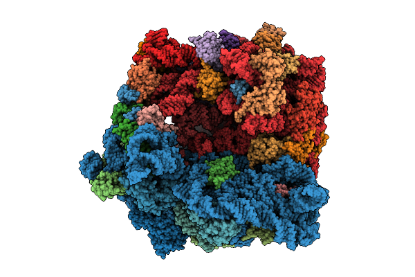 |
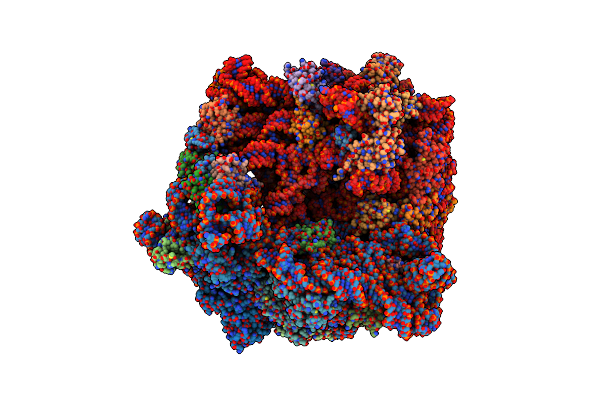
Structure Of The Bacterial Ribosome With Hypoxia-Induced Rrna Modifications
Organism: Escherichia phage t4, Escherichia coli bw25113
Method: ELECTRON MICROSCOPY Release Date: 2025-11-12 Classification: RIBOSOME Ligands: MG |
 |
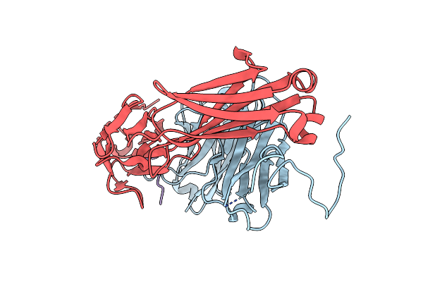
Cryo-Em Structure Of Tmprss2 In Complex With Fab Fragments Of 752 Mab And 2228 Mab
Organism: Homo sapiens, Mus musculus
Method: ELECTRON MICROSCOPY Release Date: 2025-10-15 Classification: MEMBRANE PROTEIN |
 |
Organism: Oryctolagus cuniculus, Homo sapiens
Method: X-RAY DIFFRACTION Release Date: 2025-07-02 Classification: IMMUNE SYSTEM |
 |
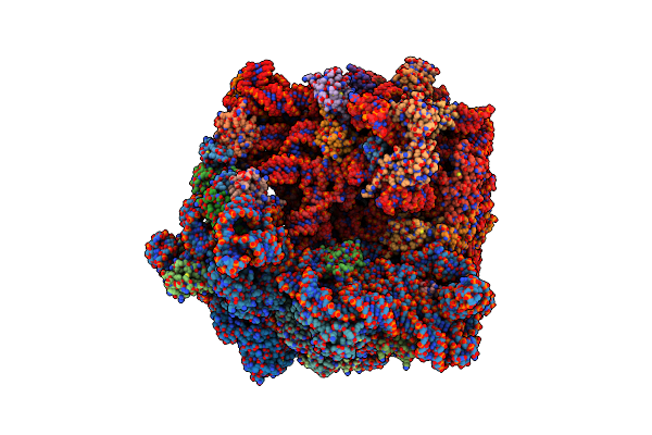
E. Coli 70S Ribosome Complexed With P. Putida Trnaile2 At The A-Site And P-Site
Organism: Escherichia coli, Pseudomonas putida nbrc 14164
Method: ELECTRON MICROSCOPY Release Date: 2024-11-06 Classification: RIBOSOME Ligands: MG |
 |
Organism: Escherichia coli, Escherichia coli bw25113, Pseudomonas putida nbrc 14164
Method: ELECTRON MICROSCOPY Release Date: 2024-11-06 Classification: RIBOSOME Ligands: MG |
 |
Organism: Escherichia coli, Escherichia coli bw25113, Pseudomonas putida nbrc 14164
Method: ELECTRON MICROSCOPY Release Date: 2024-11-06 Classification: RIBOSOME Ligands: MG |
 |
Organism: Escherichia coli, Escherichia coli bw25113, Pseudomonas putida nbrc 14164
Method: ELECTRON MICROSCOPY Release Date: 2024-11-06 Classification: RIBOSOME Ligands: MG |
 |
Organism: Escherichia coli, Escherichia coli bw25113, Pseudomonas putida nbrc 14164
Method: ELECTRON MICROSCOPY Release Date: 2024-11-06 Classification: RIBOSOME Ligands: MG |
 |
E. Coli 70S Ribosome Complexed With Trna_Ile2 Bearing L34 And T6A37 In Classical State
Organism: Escherichia coli, Escherichia coli bw25113
Method: ELECTRON MICROSCOPY Release Date: 2024-04-03 Classification: RIBOSOME Ligands: MG, K |
 |
E. Coli 70S Ribosome Complexed With H. Marismortui Trna_Ile2 Bearing Agm2C34 In Classical State
Organism: Escherichia coli, Escherichia coli bw25113, Haloarcula marismortui
Method: ELECTRON MICROSCOPY Release Date: 2024-04-03 Classification: RIBOSOME Ligands: MG, K |
 |
E. Coli 70S Ribosome Complexed With Trna_Ile2 Bearing L34 And Ct6A37 In Classical State
Organism: Escherichia coli, Escherichia coli bw25113
Method: ELECTRON MICROSCOPY Release Date: 2024-04-03 Classification: RIBOSOME Ligands: MG, K |
 |
Organism: Sulfurisphaera tokodaii
Method: X-RAY DIFFRACTION Resolution:1.90 Å Release Date: 2022-05-04 Classification: RNA |
 |
Organism: Sulfurisphaera tokodaii
Method: X-RAY DIFFRACTION Resolution:2.61 Å Release Date: 2022-05-04 Classification: RNA |
 |
Organism: Thermococcus kodakarensis
Method: X-RAY DIFFRACTION Resolution:1.80 Å Release Date: 2022-05-04 Classification: RNA BINDING PROTEIN Ligands: GMP |
 |
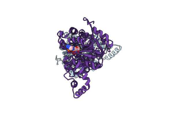
Crystal Structure Of Bacillus Subtilis Tmcal Bound With Atp (Semet Derivative)
Organism: Bacillus subtilis
Method: X-RAY DIFFRACTION Resolution:2.30 Å Release Date: 2018-07-18 Classification: LIGASE Ligands: ATP |
 |
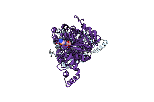
Organism: Bacillus subtilis
Method: X-RAY DIFFRACTION Resolution:2.30 Å Release Date: 2018-07-18 Classification: LIGASE |
 |
Organism: Bacillus subtilis
Method: X-RAY DIFFRACTION Resolution:2.30 Å Release Date: 2018-07-18 Classification: LIGASE Ligands: TAT |
 |
Organism: Bacillus subtilis
Method: X-RAY DIFFRACTION Resolution:2.10 Å Release Date: 2018-07-18 Classification: LIGASE Ligands: APC |
 |
Organism: Thermotoga maritima
Method: X-RAY DIFFRACTION Resolution:3.11 Å Release Date: 2018-07-18 Classification: LIGASE |