
Search Count: 15
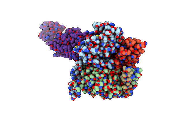 |
Organism: Homo sapiens, Rattus norvegicus, Bos taurus, Unidentified
Method: ELECTRON MICROSCOPY Release Date: 2022-08-03 Classification: SIGNALING PROTEIN |
 |
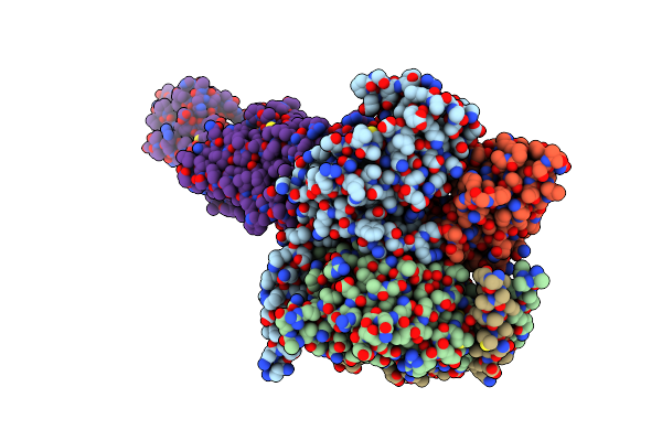
Organism: Homo sapiens, Rattus norvegicus, Bos taurus, Unidentified
Method: ELECTRON MICROSCOPY Resolution:3.30 Å Release Date: 2022-08-03 Classification: SIGNALING PROTEIN |
 |
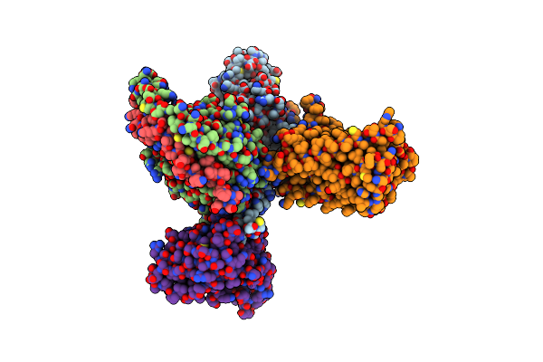
Organism: Homo sapiens, Rattus norvegicus, Bos taurus, Unidentified
Method: ELECTRON MICROSCOPY Resolution:2.80 Å Release Date: 2022-08-03 Classification: SIGNALING PROTEIN Ligands: HOH |
 |
Organism: Homo sapiens, Rattus norvegicus, Bos taurus, Unidentified
Method: ELECTRON MICROSCOPY Resolution:3.20 Å Release Date: 2022-08-03 Classification: SIGNALING PROTEIN |
 |
Organism: Homo sapiens, Rattus norvegicus, Bos taurus, Unidentified
Method: ELECTRON MICROSCOPY Resolution:3.80 Å Release Date: 2022-08-03 Classification: SIGNALING PROTEIN |
 |
Organism: Homo sapiens, Rattus norvegicus, Bos taurus, Unidentified
Method: ELECTRON MICROSCOPY Resolution:4.10 Å Release Date: 2022-08-03 Classification: SIGNALING PROTEIN |
 |
Organism: Homo sapiens, Rattus rattus, Bos taurus, Mus musculus
Method: ELECTRON MICROSCOPY Release Date: 2021-08-18 Classification: SIGNALING PROTEIN Ligands: JEV |
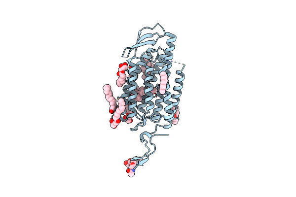 |
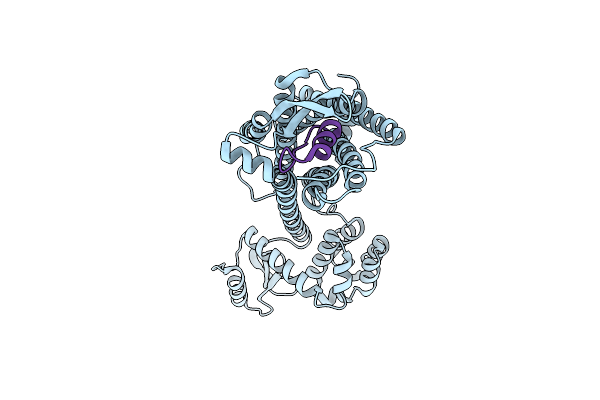
Time-Resolved Serial Femtosecond Crystallography Reveals Early Structural Changes In Channelrhodopsin: Dark State Structure
Organism: Chlamydomonas reinhardtii
Method: X-RAY DIFFRACTION Resolution:2.30 Å Release Date: 2021-04-07 Classification: MEMBRANE PROTEIN Ligands: RET, OLC |
 |
Time-Resolved Serial Femtosecond Crystallography Reveals Early Structural Changes In Channelrhodopsin: 4 Ms Structure
Organism: Chlamydomonas reinhardtii
Method: X-RAY DIFFRACTION Resolution:2.50 Å Release Date: 2021-04-07 Classification: MEMBRANE PROTEIN Ligands: RET, OLC |
 |
Time-Resolved Serial Femtosecond Crystallography Reveals Early Structural Changes In Channelrhodopsin: 1 Microsecond Structure
Organism: Chlamydomonas reinhardtii
Method: X-RAY DIFFRACTION Resolution:2.50 Å Release Date: 2021-04-07 Classification: MEMBRANE PROTEIN Ligands: RET, OLC |
 |
Time-Resolved Serial Femtosecond Crystallography Reveals Early Structural Changes In Channelrhodopsin: 50 Microsecond Structure
Organism: Chlamydomonas reinhardtii
Method: X-RAY DIFFRACTION Resolution:2.50 Å Release Date: 2021-04-07 Classification: MEMBRANE PROTEIN Ligands: RET, OLC |
 |
Time-Resolved Serial Femtosecond Crystallography Reveals Early Structural Changes In Channelrhodopsin: 250 Microsecond Structure
Organism: Chlamydomonas reinhardtii
Method: X-RAY DIFFRACTION Resolution:2.50 Å Release Date: 2021-04-07 Classification: MEMBRANE PROTEIN Ligands: RET, OLC |
 |
Time-Resolved Serial Femtosecond Crystallography Reveals Early Structural Changes In Channelrhodopsin: 1 Ms Structure
Organism: Chlamydomonas reinhardtii
Method: X-RAY DIFFRACTION Resolution:2.50 Å Release Date: 2021-04-07 Classification: MEMBRANE PROTEIN Ligands: RET, OLC |
 |
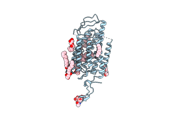
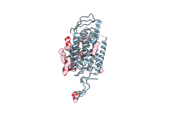
Crystal Structure Of Human Endothelin Etb Receptor In Complex With Sarafotoxin S6B
Organism: Homo sapiens, Enterobacteria phage rb59, Atractaspis engaddensis
Method: X-RAY DIFFRACTION Resolution:3.00 Å Release Date: 2020-02-12 Classification: MEMBRANE PROTEIN |
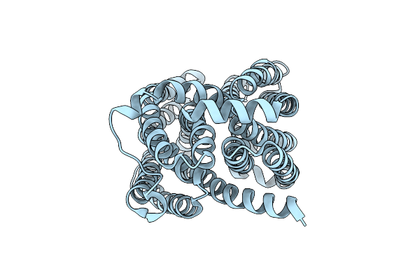 |
Organism: Arabidopsis thaliana
Method: X-RAY DIFFRACTION Resolution:2.60 Å Release Date: 2017-12-06 Classification: MEMBRANE PROTEIN |