
Search Count: 33
 |
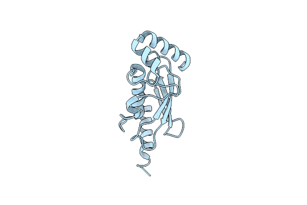
Organism: Synthetic construct
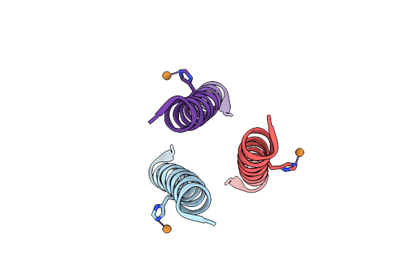
Method: X-RAY DIFFRACTION Resolution:1.45 Å Release Date: 2021-08-11 Classification: DE NOVO PROTEIN Ligands: CU |
 |
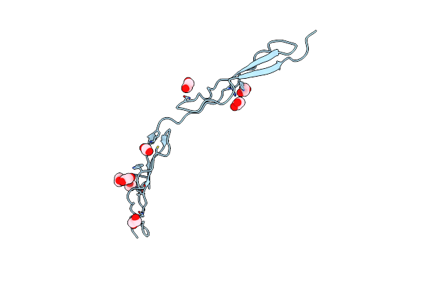
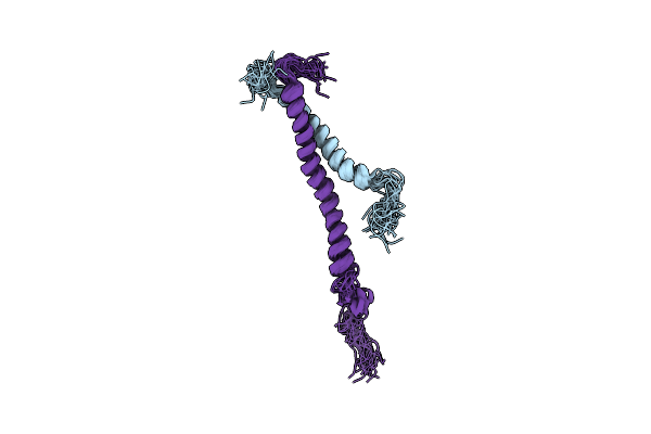
Organism: Plasmodium falciparum
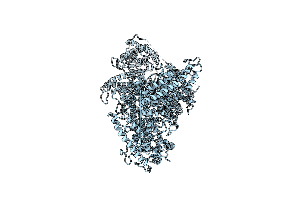
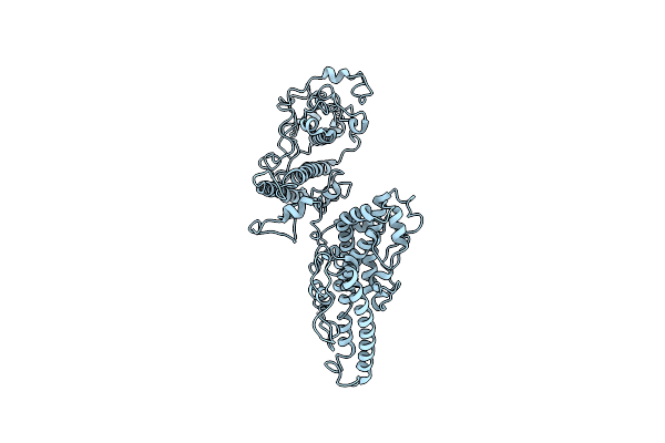
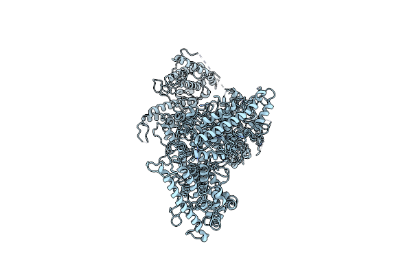
Method: ELECTRON MICROSCOPY Release Date: 2021-06-02 Classification: CELL ADHESION |
 |
Organism: Plasmodium falciparum
Method: ELECTRON MICROSCOPY Release Date: 2021-06-02 Classification: CELL ADHESION |
 |
Organism: Plasmodium falciparum
Method: ELECTRON MICROSCOPY Release Date: 2021-04-21 Classification: CELL ADHESION |
 |
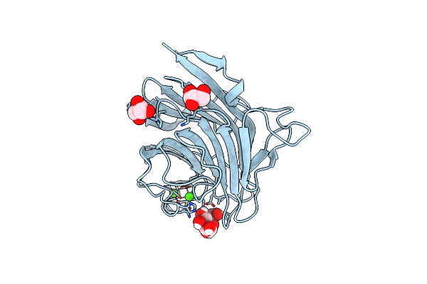
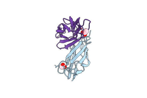
Organism: Homo sapiens
Method: X-RAY DIFFRACTION Resolution:2.46 Å Release Date: 2020-09-30 Classification: SUGAR BINDING PROTEIN Ligands: NAG |
 |
Organism: Homo sapiens
Method: X-RAY DIFFRACTION Resolution:2.54 Å Release Date: 2020-06-24 Classification: SUGAR BINDING PROTEIN |
 |
Structure Of The Chip-Tpr Domain In Complex With The Hsc70 Lid-Tail Domains
Organism: Homo sapiens
Method: X-RAY DIFFRACTION Resolution:2.91 Å Release Date: 2015-01-14 Classification: LIGASE/PROTEIN BINDING |
 |
Structural Characterization Of Inducible Nitric Oxide Synthase Substituted With Mesoheme
Organism: Mus musculus
Method: X-RAY DIFFRACTION Resolution:2.78 Å Release Date: 2014-04-23 Classification: OXIDOREDUCTASE Ligands: MH0, H4B, SO4 |
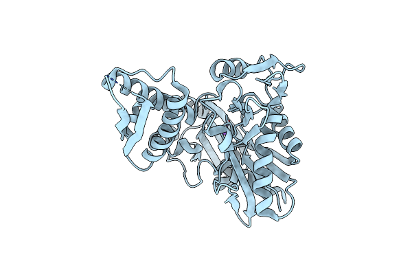 |
Crystal Structure Of The Human Mortalin (Grp75) Atpase Domain In The Apo Form
Organism: Homo sapiens
Method: X-RAY DIFFRACTION Resolution:2.80 Å Release Date: 2014-04-02 Classification: SIGNALING PROTEIN |
 |
Organism: Homo sapiens
Method: X-RAY DIFFRACTION Resolution:2.70 Å Release Date: 2013-06-05 Classification: PROTEIN TRANSPORT Ligands: CA, GOL |
 |
Organism: Homo sapiens
Method: X-RAY DIFFRACTION Resolution:2.42 Å Release Date: 2013-06-05 Classification: PROTEIN TRANSPORT Ligands: CA, MAN, GOL |
 |
Organism: Schistosoma haematobium
Method: X-RAY DIFFRACTION Resolution:2.41 Å Release Date: 2012-05-30 Classification: HYDROLASE INHIBITOR |
 |
Organism: Homo sapiens
Method: X-RAY DIFFRACTION Resolution:2.35 Å Release Date: 2012-05-16 Classification: LIGASE/PROTEIN BINDING Ligands: GOL |
 |
Organism: Homo sapiens
Method: X-RAY DIFFRACTION Resolution:1.80 Å Release Date: 2011-08-31 Classification: LIGASE |
 |
Organism: Homo sapiens
Method: X-RAY DIFFRACTION Resolution:2.40 Å Release Date: 2011-08-10 Classification: CELL ADHESION Ligands: EDO |
 |
Organism: Homo sapiens
Method: X-RAY DIFFRACTION Resolution:2.44 Å Release Date: 2011-08-03 Classification: CELL ADHESION Ligands: ACT |
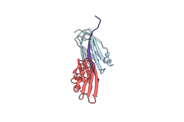 |
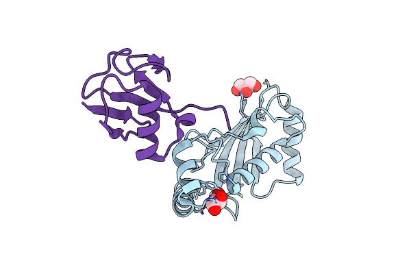
Crystal Structure Of Filamin-A Immunoglobulin-Like Repeat 21 Bound To An N-Terminal Peptide Of Cftr
Organism: Homo sapiens
Method: X-RAY DIFFRACTION Resolution:2.80 Å Release Date: 2010-04-07 Classification: STRUCTURAL PROTEIN |
 |
 |
Organism: Mus musculus
Method: X-RAY DIFFRACTION Resolution:2.30 Å Release Date: 2008-11-25 Classification: CHAPERONE Ligands: SCN, NA |
 |
Organism: Mus musculus
Method: X-RAY DIFFRACTION Resolution:2.55 Å Release Date: 2008-11-25 Classification: CHAPERONE |