
Search Count: 35
 |
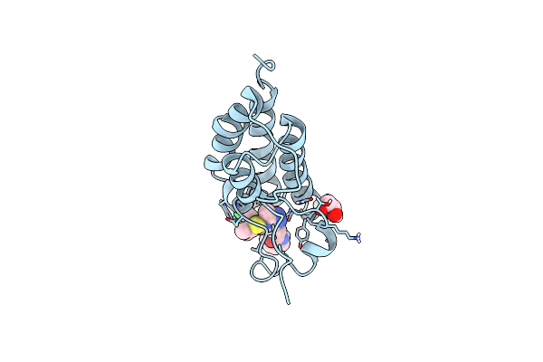
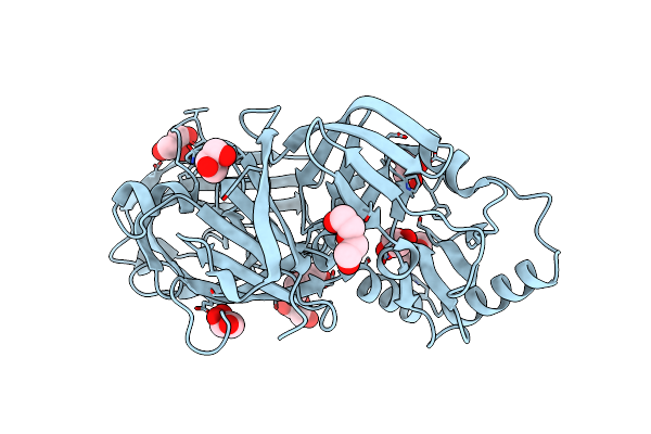
Organism: Homo sapiens
Method: X-RAY DIFFRACTION Resolution:2.72 Å Release Date: 2024-03-20 Classification: HYDROLASE Ligands: GOL |
 |
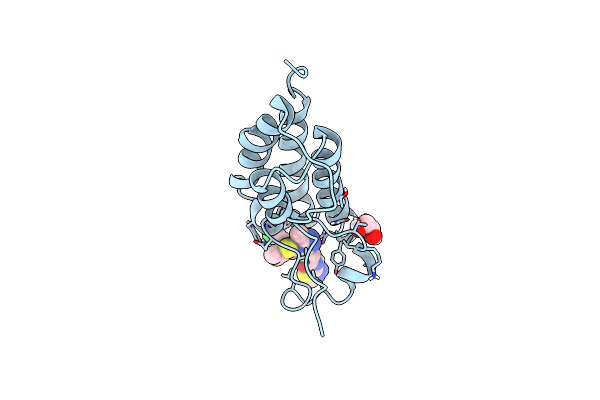
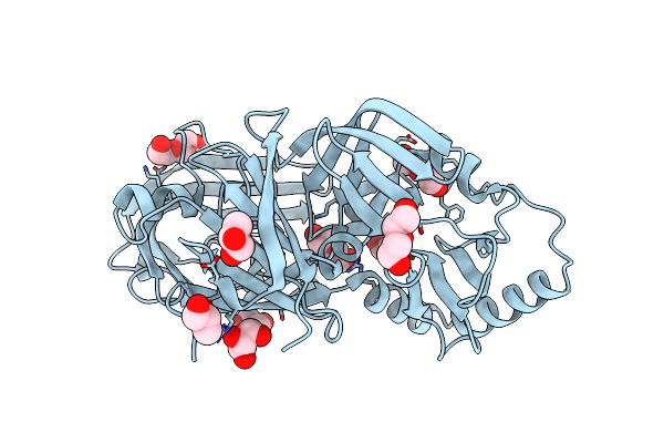
Organism: Homo sapiens
Method: X-RAY DIFFRACTION Resolution:1.85 Å Release Date: 2024-03-20 Classification: HYDROLASE Ligands: SO4 |
 |
Crystal Structure Of Fada2 (Rv0243) From The Fatty Acid Metabolic Pathway Of Mycobacterium Tuberculosis
Organism: Mycobacterium tuberculosis h37rv
Method: X-RAY DIFFRACTION Resolution:2.90 Å Release Date: 2023-04-26 Classification: TRANSFERASE Ligands: SO4 |
 |
Crystal Structure Of Ritonavir Bound Plasmepsin Ii (Pmii) From Plasmodium Falciparum
Organism: Plasmodium falciparum (isolate 3d7)
Method: X-RAY DIFFRACTION Resolution:1.90 Å Release Date: 2023-02-01 Classification: HYDROLASE Ligands: RIT, CPS, EDO |
 |
Crystal Structure Of Lopinavir Bound Plasmepsin Ii (Pmii) From Plasmodium Falciparum
Organism: Plasmodium falciparum (isolate 3d7)
Method: X-RAY DIFFRACTION Resolution:3.20 Å Release Date: 2023-02-01 Classification: HYDROLASE Ligands: AB1, CPS |
 |
Crystal Structure Of The First Bromodomain Of Human Brd4 In Complex With Sj001011461-1
Organism: Homo sapiens
Method: X-RAY DIFFRACTION Resolution:1.18 Å Release Date: 2022-08-03 Classification: CELL CYCLE Ligands: EDO, 5Z0 |
 |
Crystal Structure Of The First Bromodomain Of Human Brd4 In Complex With Sj001010551-2
Organism: Homo sapiens
Method: X-RAY DIFFRACTION Resolution:1.05 Å Release Date: 2022-08-03 Classification: CELL CYCLE Ligands: EDO, 5ZQ |
 |
Organism: Homo sapiens
Method: X-RAY DIFFRACTION Resolution:4.00 Å Release Date: 2022-06-01 Classification: HYDROLASE |
 |
Organism: Plasmodium falciparum (isolate 3d7)
Method: X-RAY DIFFRACTION Resolution:2.10 Å Release Date: 2022-02-02 Classification: HYDROLASE Ligands: NAG, EDO |
 |
Structure Of Rna-Dependent Rna Polymerase 2 (Rdr2) From Arabidopsis Thaliana
Organism: Arabidopsis thaliana
Method: ELECTRON MICROSCOPY Release Date: 2021-12-08 Classification: TRANSCRIPTION Ligands: MG |
 |
Organism: Arabidopsis thaliana
Method: ELECTRON MICROSCOPY Release Date: 2021-12-08 Classification: TRANSCRIPTION Ligands: MG |
 |
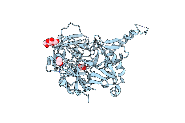
Crystal Structure Of Plasmodium Falciparum Histo-Aspartic Protease (Hap) Zymogen (Form 1)
Organism: Plasmodium falciparum
Method: X-RAY DIFFRACTION Resolution:2.00 Å Release Date: 2020-05-27 Classification: HYDROLASE Ligands: GOL, PEG |
 |
Crystal Structure Of Plasmodium Falciparum Histo-Aspartic Protease (Hap) Zymogen (Form 2)
Organism: Plasmodium falciparum
Method: X-RAY DIFFRACTION Resolution:2.50 Å Release Date: 2020-05-27 Classification: HYDROLASE Ligands: PEG, GOL |
 |
Crystal Structure Of Plasmodium Falciparum Histo-Aspartic Protease (Hap) Zymogen (Form 3)
Organism: Plasmodium falciparum
Method: X-RAY DIFFRACTION Resolution:2.90 Å Release Date: 2020-05-27 Classification: HYDROLASE Ligands: GOL |
 |
Crystal Structure Of Kni-10343 Bound Plasmepsin Ii (Pmii) From Plasmodium Falciparum
Organism: Plasmodium falciparum
Method: X-RAY DIFFRACTION Resolution:2.00 Å Release Date: 2018-07-11 Classification: HYDROLASE Ligands: 8V9, CPS, GOL, PO4 |
 |
Crystal Structure Of Kni-10743 Bound Plasmepsin Ii (Pmii) From Plasmodium Falciparum
Organism: Plasmodium falciparum
Method: X-RAY DIFFRACTION Resolution:2.15 Å Release Date: 2018-07-11 Classification: HYDROLASE Ligands: 8VC, GOL, EDO, CPS |
 |
Crystal Structure Of Kni-10333 Bound Plasmepsin Ii (Pmii) From Plasmodium Falciparum
Organism: Plasmodium falciparum
Method: X-RAY DIFFRACTION Resolution:1.90 Å Release Date: 2018-07-11 Classification: HYDROLASE Ligands: 8VO, CPS, GOL |
 |
Crystal Structure Of Kni-10395 Bound Plasmepsin Ii (Pmii) From Plasmodium Falciparum
Organism: Plasmodium falciparum
Method: X-RAY DIFFRACTION Resolution:2.10 Å Release Date: 2018-07-11 Classification: HYDROLASE Ligands: K95, CPS, NA |
 |
Crystal Structure Of Kni-10742 Bound Plasmepsin Ii (Pmii) From Plasmodium Falciparum
Organism: Plasmodium falciparum (isolate 3d7)
Method: X-RAY DIFFRACTION Resolution:2.10 Å Release Date: 2018-07-11 Classification: HYDROLASE Ligands: CPS, 8VF, NA |
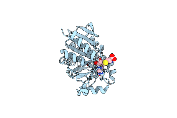 |
Organism: Helicobacter pylori (strain j99 / atcc 700824)
Method: X-RAY DIFFRACTION, NEUTRON DIFFRACTION Release Date: 2016-11-23 Classification: HYDROLASE Ligands: SAH, DOD |