
Search Count: 7
 |
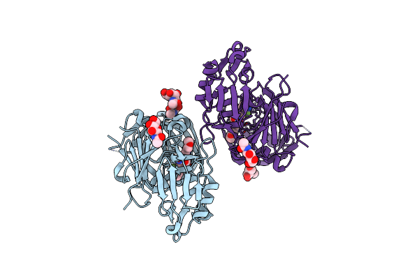
Crystal Structure Of Human Kras G12C Covalently Bound To A Phthalazine Inhibitor
Organism: Homo sapiens
Method: X-RAY DIFFRACTION Resolution:1.60 Å Release Date: 2019-12-25 Classification: SIGNALING PROTEIN/Inhibitor Ligands: MG, GDP, OJ1 |
 |
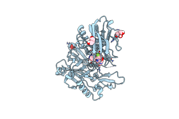
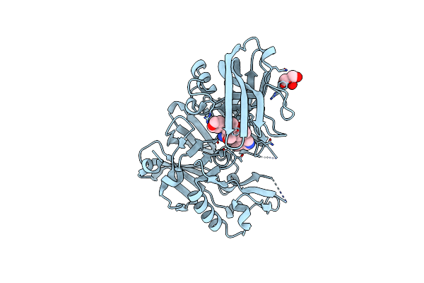
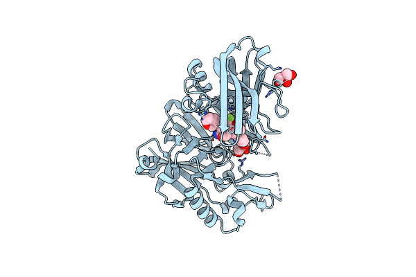
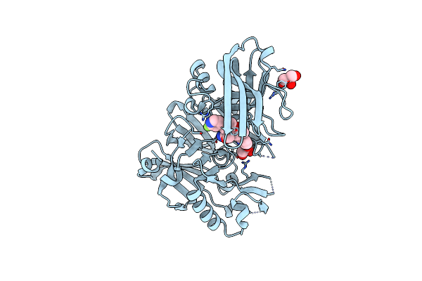
Crystal Structure Of Human Kras G12C Covalently Bound To A Quinazolinone Inhibitor
Organism: Homo sapiens
Method: X-RAY DIFFRACTION Resolution:1.50 Å Release Date: 2019-12-25 Classification: SIGNALING PROTEIN/Inhibitor Ligands: GDP, CA, OHY |
 |
Crystal Structure Of Rat Cathepsin D With (5S)-3-(5,6-Dihydro-2H-Pyran-3-Yl)-1-Fluoro- 7-(2-Fluoropyridin-3-Yl)Spiro[Chromeno[2,3- C]Pyridine-5,4'-[1,3]Oxazol]-2'-Amine
Organism: Rattus norvegicus
Method: X-RAY DIFFRACTION Resolution:2.81 Å Release Date: 2018-06-13 Classification: HYDROLASE Ligands: NAG, 3UT |
 |
Structure Of Bace-1 (Beta-Secretase) In Complex With : N-(3-((1R,5S,6R)-3-Amino-5-Methyl-2-Oxa-4-Azabicyclo[4.1.0]Hept-3-En-5-Yl)-4-Fluorophenyl)-5-Methoxypyrazine-2-Carboxamide
Organism: Homo sapiens
Method: X-RAY DIFFRACTION Resolution:1.95 Å Release Date: 2018-02-21 Classification: Hydrolase/Hydrolase Inhibitor Ligands: IOD, GOL, SO4, EJ7 |
 |
Crystal Structure Of Bace1 In Complex With 2-Aminooxazoline-3-Azaxanthene Compound 12
Organism: Homo sapiens
Method: X-RAY DIFFRACTION Resolution:1.90 Å Release Date: 2017-05-17 Classification: HYDROLASE Ligands: IOD, 8QV, GOL |
 |
Crystal Structure Of Bace1 In Complex With 2-Aminooxazoline 3-Aza-4-Fluoro-Xanthene Inhibitor 22
Organism: Homo sapiens
Method: X-RAY DIFFRACTION Resolution:1.85 Å Release Date: 2015-03-04 Classification: Hydrolase/Hydrolase Inhibitor Ligands: IOD, GOL, 3UT |
 |
Crystal Structure Of Bace1 In Complex With 2-Aminooxazoline 3-Azaxanthene Inhibitor 28
Organism: Homo sapiens
Method: X-RAY DIFFRACTION Resolution:1.80 Å Release Date: 2015-02-04 Classification: Hydrolase/hydrolase inhibitor Ligands: IOD, 43K, GOL |