
Search Count: 498
 |
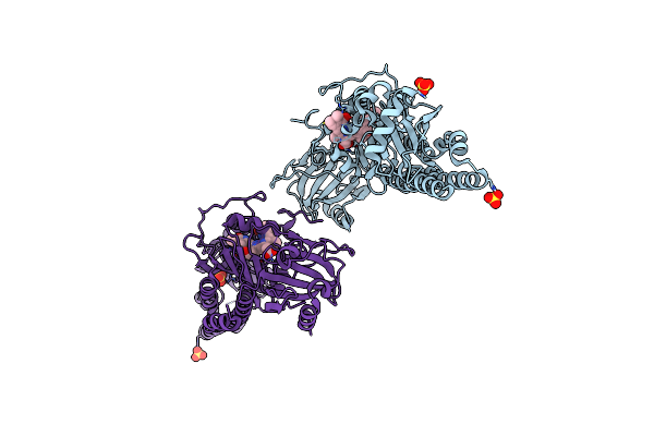
Organism: Sars-cov-2 pseudovirus
Method: X-RAY DIFFRACTION Resolution:2.38 Å Release Date: 2025-09-17 Classification: VIRAL PROTEIN Ligands: ZN, SAH, A1JLH, CL, IMD |
 |
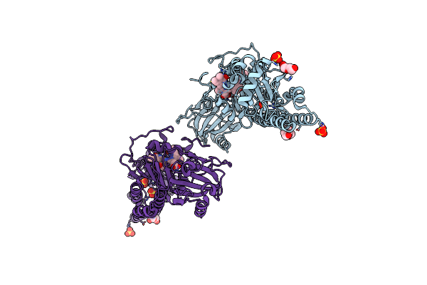
Organism: Severe acute respiratory syndrome coronavirus
Method: X-RAY DIFFRACTION Resolution:1.89 Å Release Date: 2025-08-27 Classification: VIRAL PROTEIN Ligands: ZN, SAH, A1JDF, CL, EDO, IMD, IPA, SO4 |
 |
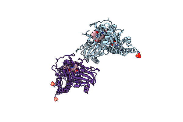
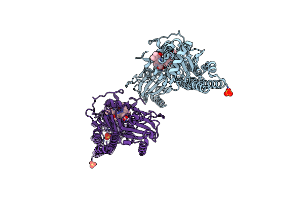
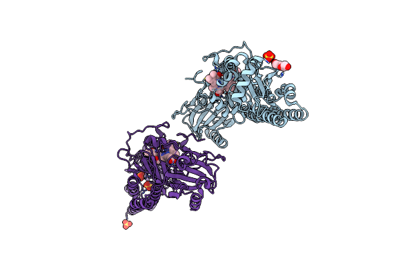
Organism: Homo sapiens, Synthetic construct
Method: X-RAY DIFFRACTION Resolution:2.86 Å Release Date: 2025-07-16 Classification: APOPTOSIS |
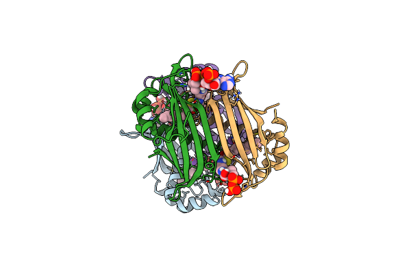 |
Crystal Structure Of The Espe7 Thioesterase Mutant R35A From The Esperamicin Biosynthetic Pathway At 1.6 A
Organism: Actinomadura verrucosospora
Method: X-RAY DIFFRACTION Resolution:1.57 Å Release Date: 2025-06-25 Classification: HYDROLASE Ligands: K, DCC |
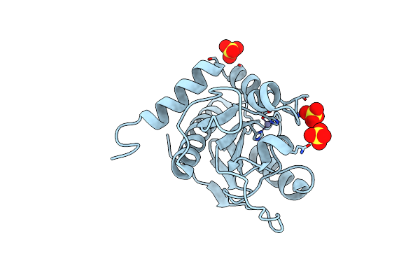 |
Organism: Listeria phage lp-125
Method: X-RAY DIFFRACTION Resolution:1.80 Å Release Date: 2025-06-25 Classification: HYDROLASE Ligands: ZN, SO4 |
 |
Serial Femtosecond X-Ray Structure Of A Fluorescence Optimized Bathy Phytochrome Pairfp2 Derived From Wild-Type Agp2 In Its Pfr State (I0A).
Organism: Agrobacterium fabrum str. c58
Method: X-RAY DIFFRACTION Resolution:2.15 Å Release Date: 2025-05-14 Classification: SIGNALING PROTEIN Ligands: EL5, SO4, CL, EDO |
 |
Serial Femtosecond X-Ray Structure Of A Fluorescence Optimized Bathy Phytochrome Pairfp2 Derived From Wild-Type Agp2 In Its Pfr State (I0B).
Organism: Agrobacterium fabrum str. c58
Method: X-RAY DIFFRACTION Resolution:2.20 Å Release Date: 2025-05-14 Classification: SIGNALING PROTEIN Ligands: EL5, SO4 |
 |
Serial Femtosecond X-Ray Structure Of A Fluorescence Optimized Bathy Phytochrome Pairfp2 Derived From Wild-Type Agp2 In I1 Intermediate State.
Organism: Agrobacterium fabrum str. c58
Method: X-RAY DIFFRACTION Resolution:2.54 Å Release Date: 2025-05-14 Classification: SIGNALING PROTEIN Ligands: EL5, SO4 |
 |
Serial Femtosecond X-Ray Structure Of A Fluorescence Optimized Bathy Phytochrome Pairfp2 Derived From Wild-Type Agp2 In I2 Intermediate State.
Organism: Agrobacterium fabrum str. c58
Method: X-RAY DIFFRACTION Resolution:2.43 Å Release Date: 2025-05-14 Classification: SIGNALING PROTEIN Ligands: EL5, SO4, PGE, PEG, CL |
 |
Serial Femtosecond X-Ray Structure Of A Fluorescence Optimized Bathy Phytochrome Pairfp2 Derived From Wild-Type Agp2 In I3 Intermediate State.
Organism: Agrobacterium fabrum str. c58
Method: X-RAY DIFFRACTION Resolution:2.40 Å Release Date: 2025-05-14 Classification: SIGNALING PROTEIN Ligands: EL5, SO4, GOL, PEG |
 |
Serial Femtosecond X-Ray Structure Of A Fluorescence Optimized Bathy Phytochrome Pairfp2 Derived From Wild-Type Agp2 In I4 Intermediate State.
Organism: Agrobacterium fabrum str. c58
Method: X-RAY DIFFRACTION Resolution:2.30 Å Release Date: 2025-05-14 Classification: SIGNALING PROTEIN Ligands: EL5, SO4, CL, PEG |
 |
Serial Femtosecond X-Ray Structure Of A Fluorescence Optimized Bathy Phytochrome Pairfp2 Derived From Wild-Type Agp2 In I5 Intermediate State.
Organism: Agrobacterium fabrum str. c58
Method: X-RAY DIFFRACTION Resolution:2.43 Å Release Date: 2025-05-14 Classification: SIGNALING PROTEIN Ligands: EL5, SO4, CL |
 |
Serial Femtosecond X-Ray Structure Of A Fluorescence Optimized Bathy Phytochrome Pairfp2 Derived From Wild-Type Agp2 In I6 Intermediate State.
Organism: Agrobacterium fabrum str. c58
Method: X-RAY DIFFRACTION Resolution:2.49 Å Release Date: 2025-05-14 Classification: SIGNALING PROTEIN Ligands: EL5, SO4, CL |
 |
Serial Femtosecond X-Ray Structure Of A Fluorescence Optimized Bathy Phytochrome Pairfp2 Derived From Wild-Type Agp2 In I7 Intermediate State.
Organism: Agrobacterium fabrum str. c58
Method: X-RAY DIFFRACTION Resolution:2.80 Å Release Date: 2025-05-14 Classification: SIGNALING PROTEIN Ligands: EL5, SO4, PEG |
 |
Structure Of E. Coli Dihydrofolate Reductase (Dhfr) In An Occluded Conformation And In Complex With A Cycloguanil Derivative
Organism: Escherichia coli
Method: X-RAY DIFFRACTION Resolution:2.35 Å Release Date: 2025-02-26 Classification: OXIDOREDUCTASE Ligands: A1A3E |
 |
Structure Of E. Coli Dihydrofolate Reductase (Dhfr) In An Occluded Conformation And In Complex With Cycloguanil
Organism: Escherichia coli
Method: X-RAY DIFFRACTION Resolution:2.17 Å Release Date: 2025-02-26 Classification: OXIDOREDUCTASE Ligands: 1CY, FLC |
 |
Design And Application Of Synthetic 17B-Hsd13 Substrates To Drug Discovery, And To Reveal Preserved Catalytic Activity Of Protective Human Variants
Organism: Homo sapiens
Method: X-RAY DIFFRACTION Resolution:2.09 Å Release Date: 2024-11-27 Classification: HYDROLASE Ligands: NAD, A1AG4 |
 |
Design And Application Of Synthetic 17B-Hsd13 Substrates To Drug Discovery, And To Reveal Preserved Catalytic Activity Of Protective Human Variants
Organism: Homo sapiens
Method: X-RAY DIFFRACTION Resolution:2.36 Å Release Date: 2024-11-27 Classification: HYDROLASE Ligands: NAD, A1AG5 |
 |
Design And Application Of Synthetic 17B-Hsd13 Substrates To Drug Discovery, And To Reveal Preserved Catalytic Activity Of Protective Human Variants
Organism: Homo sapiens
Method: X-RAY DIFFRACTION Resolution:2.59 Å Release Date: 2024-11-27 Classification: HYDROLASE Ligands: NAD, A1AG6 |
 |
Organism: Odocoileus virginianus
Method: ELECTRON MICROSCOPY Release Date: 2024-11-06 Classification: PROTEIN FIBRIL |