
Search Count: 24
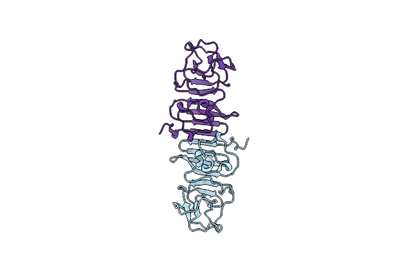 |
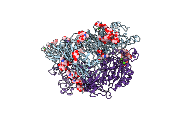
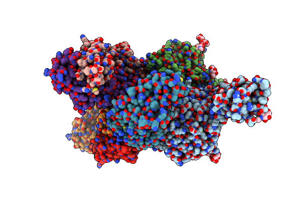
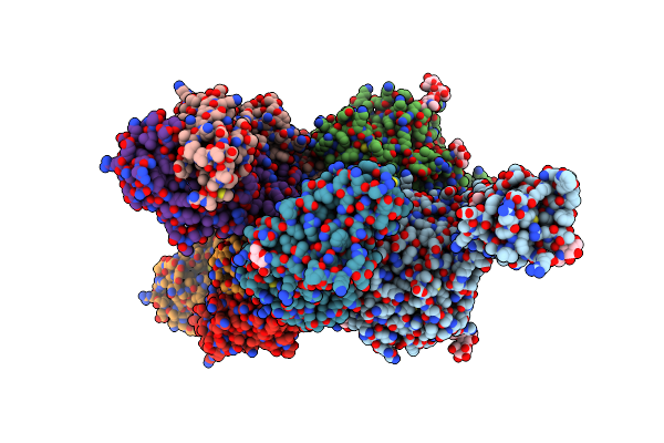
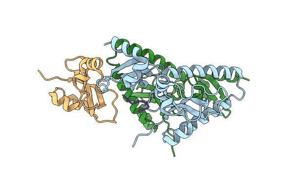
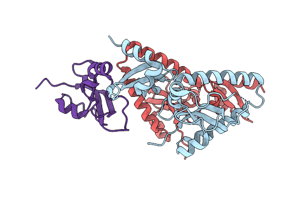
A Mutation Identified In Neonatal Microcephaly Destabilizes Zika Virus Ns1 Assembly In Vitro
Organism: Zika virus
Method: X-RAY DIFFRACTION Resolution:2.82 Å Release Date: 2017-05-17 Classification: VIRAL PROTEIN |
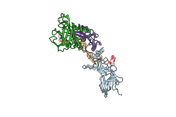 |
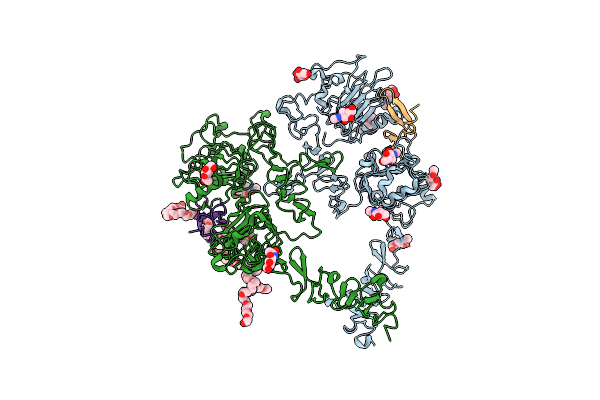
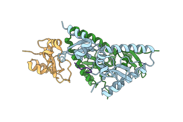
Organism: Mus musculus, Homo sapiens
Method: X-RAY DIFFRACTION Resolution:3.30 Å Release Date: 2015-03-04 Classification: CYTOKINE Ligands: ZN |
 |
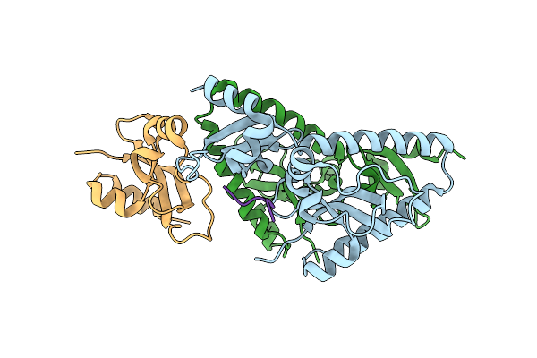
Organism: Mus musculus, Homo sapiens
Method: X-RAY DIFFRACTION Resolution:3.25 Å Release Date: 2015-03-04 Classification: CYTOKINE Ligands: NAG, ZN |
 |
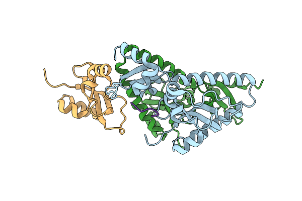
Organism: Homo sapiens
Method: X-RAY DIFFRACTION Resolution:3.00 Å Release Date: 2012-12-12 Classification: PROTEIN BINDING Ligands: CA, NAG, SO4, CL, NI |
 |
Organism: Homo sapiens
Method: X-RAY DIFFRACTION Resolution:2.90 Å Release Date: 2012-12-12 Classification: PROTEIN BINDING Ligands: CA, NAG, NA |
 |
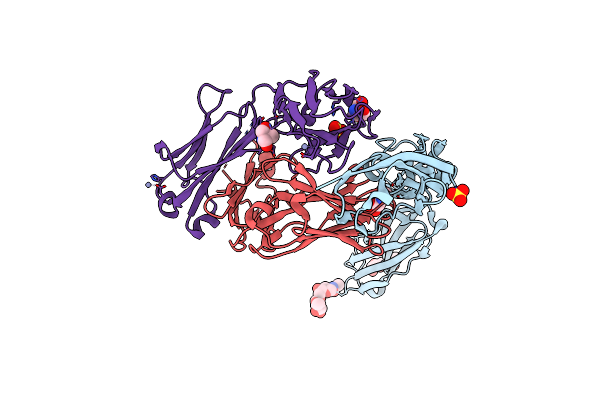
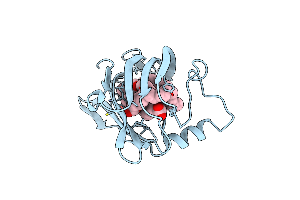
Organism: Plasmodium falciparum
Method: X-RAY DIFFRACTION Resolution:1.70 Å Release Date: 2012-05-09 Classification: CELL INVASION |
 |
Crystal Structure Of Circumsporozoite Protein Atsr Domain, R32 Platinum-Bound Form
Organism: Plasmodium falciparum
Method: X-RAY DIFFRACTION Resolution:1.85 Å Release Date: 2012-05-09 Classification: CELL INVASION Ligands: PT |
 |
Organism: Plasmodium falciparum
Method: X-RAY DIFFRACTION Resolution:2.04 Å Release Date: 2012-05-09 Classification: CELL INVASION |
 |
Organism: Homo sapiens, Mus musculus
Method: X-RAY DIFFRACTION Resolution:3.15 Å Release Date: 2012-01-11 Classification: CELL ADHESION Ligands: CA, NAG, 15P, MG, TRS |
 |
Organism: Homo sapiens, Mus musculus
Method: X-RAY DIFFRACTION Resolution:3.10 Å Release Date: 2012-01-11 Classification: CELL ADHESION Ligands: CA, NAG, TRS, MG, 0DU |
 |
The Extracellular And Transmembrane Domain Interfaces In Epidermal Growth Factor Receptor Signaling
Organism: Homo sapiens
Method: X-RAY DIFFRACTION Resolution:3.30 Å Release Date: 2010-10-13 Classification: TRANSFERASE Ligands: NAG, 2PE |
 |
Structural Plasticity In Igsf Domain 4 Of Icam-1 Mediates Cell Surface Dimerization
Organism: Homo sapiens, Mus musculus
Method: X-RAY DIFFRACTION Resolution:2.70 Å Release Date: 2007-10-16 Classification: CELL ADHESION Ligands: NAG, SO4, ZN, TRS |
 |
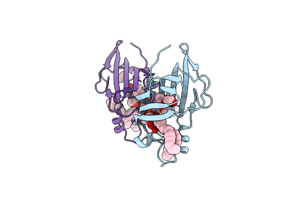
Tandem Chromodomains Of Human Chd1 Complexed With Histone H3 Tail Containing Trimethyllysine 4 And Phosphothreonine 3
Organism: Homo sapiens
Method: X-RAY DIFFRACTION Resolution:2.45 Å Release Date: 2005-12-27 Classification: PEPTIDE BINDING PROTEIN |
 |
Tandem Chromodomains Of Human Chd1 Complexed With Histone H3 Tail Containing Trimethyllysine 4 And Dimethylarginine 2
Organism: Homo sapiens
Method: X-RAY DIFFRACTION Resolution:2.95 Å Release Date: 2005-12-27 Classification: PEPTIDE BINDING PROTEIN |
 |
Crystal Structure Analysis Of Human Chd1 Chromodomains 1 And 2 Bound To Histone H3 Resi 1-15 Mek4
Organism: Homo sapiens
Method: X-RAY DIFFRACTION Resolution:2.65 Å Release Date: 2005-12-27 Classification: PEPTIDE BINDING PROTEIN |
 |
Tandem Chromodomains Of Human Chd1 Complexed With Histone H3 Tail Containing Trimethyllysine 4
Organism: Homo sapiens
Method: X-RAY DIFFRACTION Resolution:2.40 Å Release Date: 2005-12-27 Classification: PEPTIDE BINDING PROTEIN |
 |
Organism: Homo sapiens
Method: X-RAY DIFFRACTION Resolution:2.35 Å Release Date: 2005-12-27 Classification: PEPTIDE BINDING PROTEIN |
 |
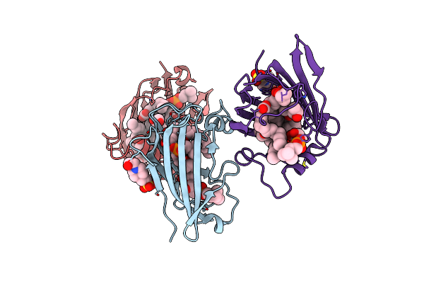
Crystal Structure Analysis Of Gm2-Activator Protein Complexed With Phosphatidylcholine
Organism: Homo sapiens
Method: X-RAY DIFFRACTION Resolution:2.00 Å Release Date: 2005-10-25 Classification: LIPID BINDING PROTEIN Ligands: OLA, MYR, DAO, IPA |
 |
Crystal Structure Analysis Of Gm2-Activator Protein Complexed With Phosphatidylcholine
Organism: Homo sapiens
Method: X-RAY DIFFRACTION Resolution:2.00 Å Release Date: 2005-10-25 Classification: LIPID BINDING PROTEIN Ligands: CL, EPE, LP3, IPA, CH5, OLA, MYR, DAO |
 |
Crystal Structure Analysis Of Gm2-Activator Protein Complexed With Phosphatidylcholine
Organism: Homo sapiens
Method: X-RAY DIFFRACTION Resolution:1.80 Å Release Date: 2005-10-25 Classification: LIPID BINDING PROTEIN Ligands: LP3, OLA, IPA |