
Search Count: 59
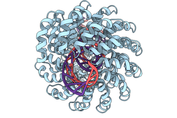 |
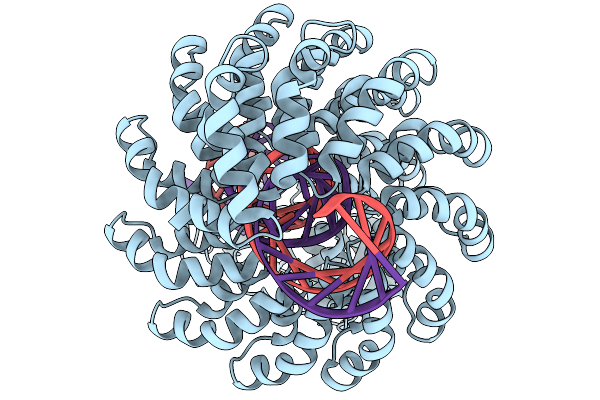
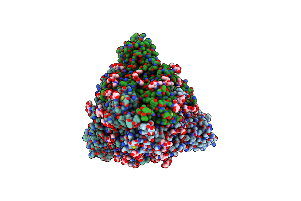
Organism: Burkholderia cenocepacia, Synthetic construct
Method: ELECTRON MICROSCOPY Release Date: 2025-11-19 Classification: DE NOVO PROTEIN Ligands: ZN |
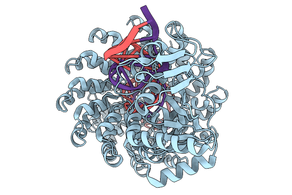 |
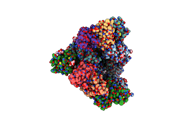
Organism: Synthetic construct
Method: ELECTRON MICROSCOPY Release Date: 2025-11-19 Classification: DE NOVO PROTEIN Ligands: ZN |
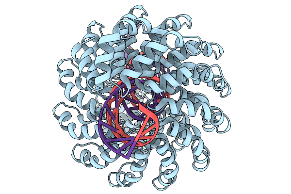 |
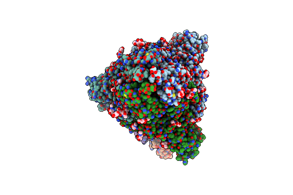
Organism: Xanthomonas, Synthetic construct
Method: ELECTRON MICROSCOPY Release Date: 2025-11-19 Classification: DE NOVO PROTEIN Ligands: ZN |
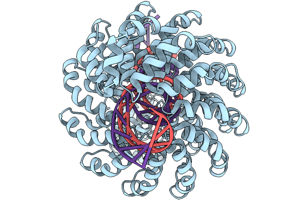 |
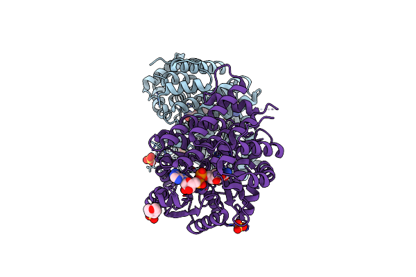
Organism: Synthetic construct
Method: ELECTRON MICROSCOPY Release Date: 2025-11-19 Classification: DE NOVO PROTEIN Ligands: ZN |
 |
Organism: Synthetic construct
Method: ELECTRON MICROSCOPY Release Date: 2025-11-19 Classification: DE NOVO PROTEIN Ligands: ZN |
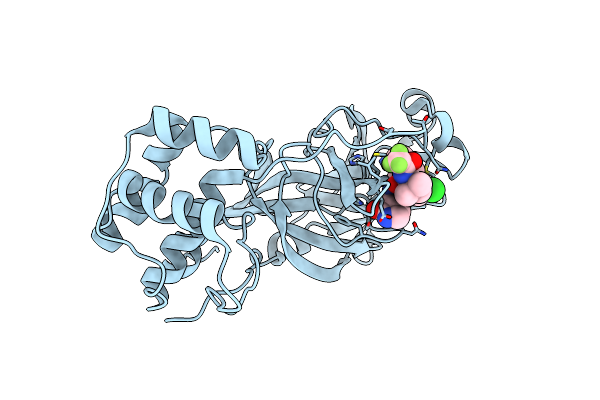 |
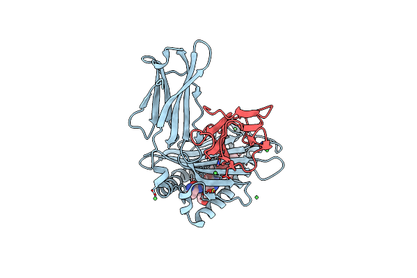
Crystal Structure Of Sars-Cov-2 Main Protease In Complex With An Inhibitor Tkb-277-5Cl
Organism: Severe acute respiratory syndrome coronavirus 2
Method: X-RAY DIFFRACTION Resolution:1.80 Å Release Date: 2025-06-04 Classification: VIRAL PROTEIN Ligands: A1B7C |
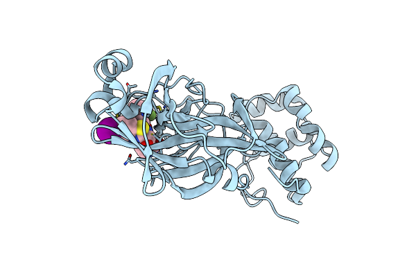 |
Crystal Structure Of Sars-Cov-2 Main Protease In Complex With An Inhibitor Tkb-280-5I
Organism: Severe acute respiratory syndrome coronavirus
Method: X-RAY DIFFRACTION Resolution:2.01 Å Release Date: 2025-06-04 Classification: VIRAL PROTEIN Ligands: A1B7A |
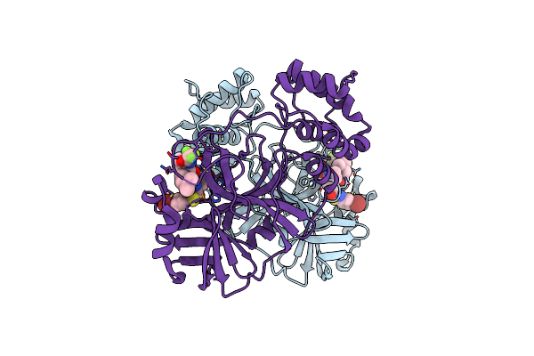 |
Crystal Structure Of Sars-Cov-2 Main Protease In Complex With An Inhibitor Tkb-276-5Br
Organism: Severe acute respiratory syndrome coronavirus 2
Method: X-RAY DIFFRACTION Resolution:1.79 Å Release Date: 2025-06-04 Classification: VIRAL PROTEIN Ligands: A1B7B |
 |
Organism: Mus musculus
Method: X-RAY DIFFRACTION Resolution:2.43 Å Release Date: 2024-06-12 Classification: IMMUNE SYSTEM Ligands: NI |
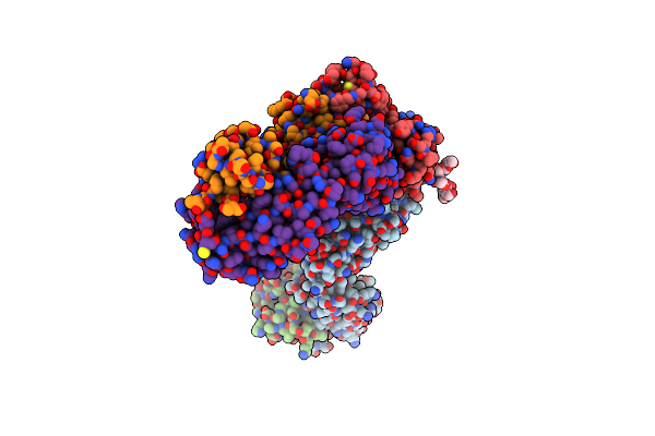 |
Structure Of The Sars-Cov-2 Rbd In Complex With Neutralizing Antibodies Bg4-25 And Cr3022
Organism: Homo sapiens, Severe acute respiratory syndrome coronavirus 2
Method: X-RAY DIFFRACTION Resolution:3.10 Å Release Date: 2021-05-05 Classification: VIRAL PROTEIN/ANTIVIRAL PROTEIN |
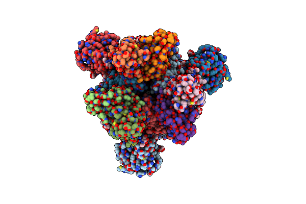 |
Structure Of The Sars-Cov-2 S 6P Trimer In Complex With The Human Neutralizing Antibody Fab Fragment, Bg10-19
Organism: Severe acute respiratory syndrome coronavirus 2, Homo sapiens
Method: ELECTRON MICROSCOPY Release Date: 2021-05-05 Classification: VIRAL PROTEIN/ANTIVIRAL PROTEIN Ligands: NAG |
 |
Structure Of The Sars-Cov-2 S 6P Trimer In Complex With The Human Neutralizing Antibody Fab Fragment, Bg1-22
Organism: Severe acute respiratory syndrome coronavirus 2, Homo sapiens
Method: ELECTRON MICROSCOPY Release Date: 2021-05-05 Classification: VIRAL PROTEIN/ANTIVIRAL PROTEIN Ligands: NAG |
 |
Structure Of The Sars-Cov-2 S 6P Trimer In Complex With The Human Neutralizing Antibody Fab Fragment, Bg7-15
Organism: Severe acute respiratory syndrome coronavirus 2, Homo sapiens
Method: ELECTRON MICROSCOPY Release Date: 2021-05-05 Classification: VIRAL PROTEIN/ANTIVIRAL PROTEIN Ligands: NAG |
 |
Structure Of The Sars-Cov-2 S 2P Trimer In Complex With The Human Neutralizing Antibody Fab Fragment, Bg7-20
Organism: Severe acute respiratory syndrome coronavirus 2, Homo sapiens
Method: ELECTRON MICROSCOPY Release Date: 2021-05-05 Classification: VIRAL PROTEIN/ANTIVIRAL PROTEIN Ligands: NAG |
 |
Structure Of The Sars-Cov-2 S 2P Trimer In Complex With The Human Neutralizing Antibody Fab Fragment, Bg1-24
Organism: Severe acute respiratory syndrome coronavirus 2, Homo sapiens
Method: ELECTRON MICROSCOPY Release Date: 2021-05-05 Classification: VIRAL PROTEIN/ANTIVIRAL PROTEIN Ligands: NAG |
 |
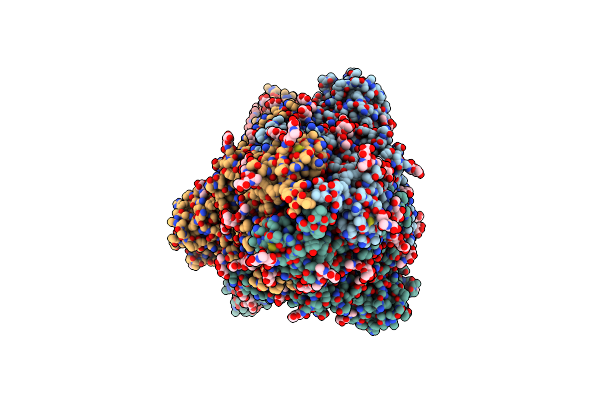
Organism: Escherichia coli (strain k12)
Method: X-RAY DIFFRACTION Resolution:1.65 Å Release Date: 2020-08-26 Classification: OXIDOREDUCTASE Ligands: NAD, SO4, EDO, FE, MES |
 |
Organism: Escherichia coli (strain k12)
Method: X-RAY DIFFRACTION Resolution:1.95 Å Release Date: 2020-08-26 Classification: OXIDOREDUCTASE Ligands: FE |
 |
 |
Organism: Homo sapiens
Method: X-RAY DIFFRACTION Resolution:2.10 Å Release Date: 2020-02-05 Classification: HYDROLASE Ligands: J1W |
 |
Organism: Homo sapiens
Method: X-RAY DIFFRACTION Resolution:2.60 Å Release Date: 2020-02-05 Classification: HYDROLASE Ligands: PLM |