
Search Count: 107
 |
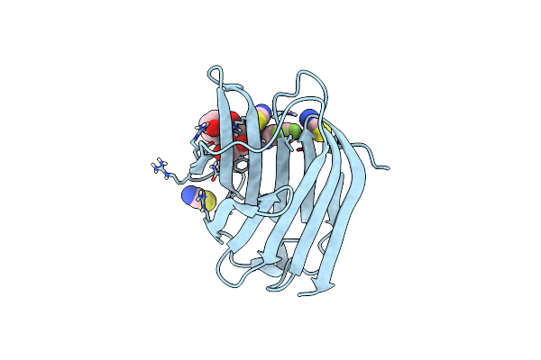
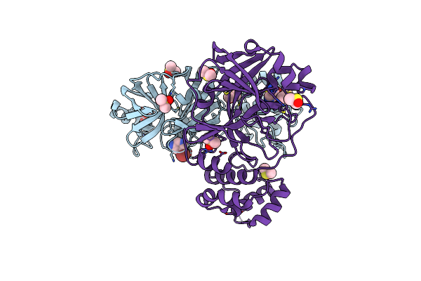
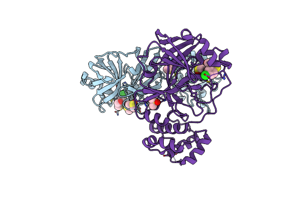
Cdk2/Cycline Bound To Compound 11 With P-Loop In The Ee And Cc Conformations
Organism: Homo sapiens
Method: X-RAY DIFFRACTION Resolution:2.12 Å Release Date: 2025-04-30 Classification: CELL CYCLE Ligands: A1CAE |
 |
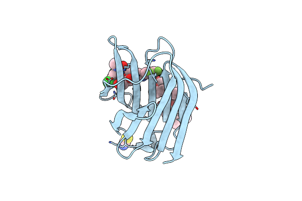
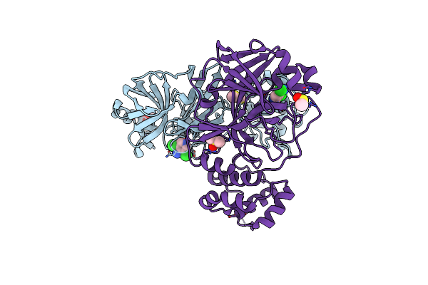
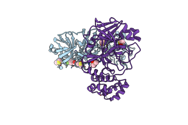
Organism: Homo sapiens
Method: X-RAY DIFFRACTION Resolution:1.98 Å Release Date: 2025-04-30 Classification: CELL CYCLE Ligands: A1CAH |
 |
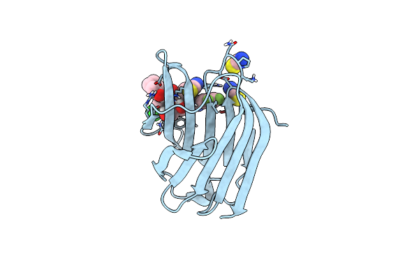
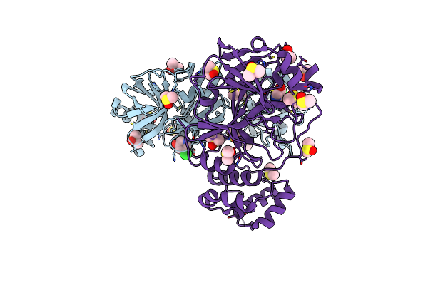
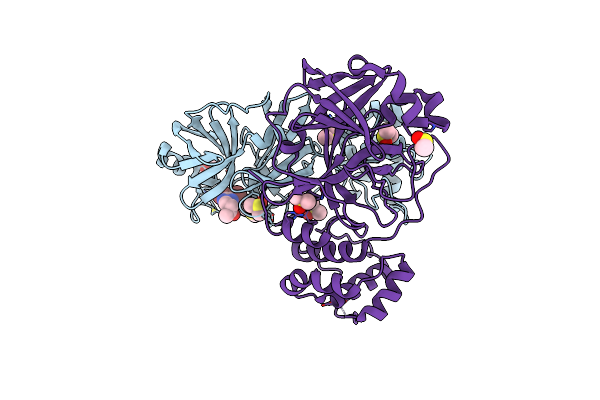
Cdk2/Cycline Bound To Compound 19 With P-Loop In The Ee And Ec Conformations
Organism: Homo sapiens
Method: X-RAY DIFFRACTION Resolution:1.95 Å Release Date: 2025-04-30 Classification: CELL CYCLE Ligands: A1CAF |
 |
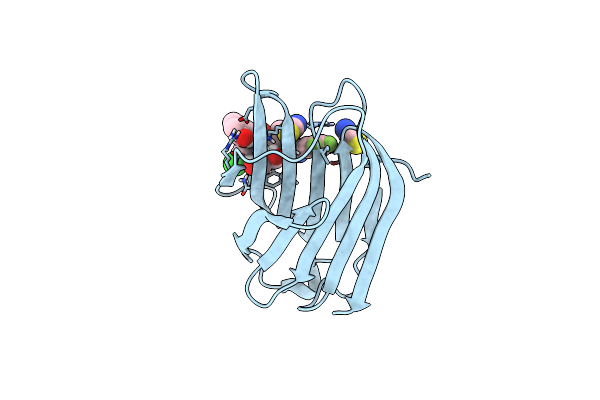
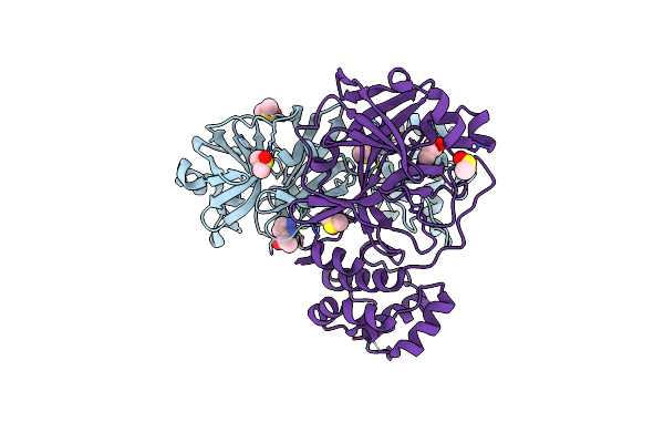
Cdk2/Cycline Bound To Compound 20 With P-Loop In The Ee And Cc Conformations
Organism: Homo sapiens
Method: X-RAY DIFFRACTION Resolution:2.10 Å Release Date: 2025-04-30 Classification: CELL CYCLE Ligands: A1CAG |
 |
Cdk2/Cycline Bound To Compound 21 With P-Loop In The Ee And Cc Conformations
Organism: Homo sapiens
Method: X-RAY DIFFRACTION Resolution:2.00 Å Release Date: 2025-04-30 Classification: CELL CYCLE Ligands: A1CAI |
 |
Organism: Homo sapiens
Method: X-RAY DIFFRACTION Release Date: 2025-03-26 Classification: SUGAR BINDING PROTEIN Ligands: SCN, A1IB3 |
 |
Organism: Homo sapiens
Method: X-RAY DIFFRACTION Release Date: 2025-03-26 Classification: SUGAR BINDING PROTEIN Ligands: 2PE, A1IB4, SCN |
 |
Organism: Homo sapiens
Method: X-RAY DIFFRACTION Release Date: 2025-01-29 Classification: SUGAR BINDING PROTEIN Ligands: A1H1W, SCN |
 |
Organism: Homo sapiens
Method: X-RAY DIFFRACTION Release Date: 2025-01-29 Classification: SUGAR BINDING PROTEIN Ligands: A1H1Y, SCN |
 |
Organism: Homo sapiens
Method: X-RAY DIFFRACTION Release Date: 2025-01-29 Classification: SUGAR BINDING PROTEIN Ligands: A1H1X, SCN |
 |
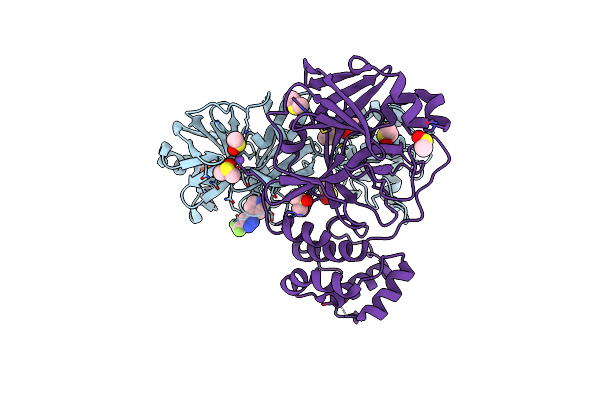
Macrocyclic Inhibitors Targeting The Prime Site Of The Fibrinolytic Serine Protease Plasmin
Organism: Homo sapiens
Method: X-RAY DIFFRACTION Resolution:2.10 Å Release Date: 2024-10-30 Classification: HYDROLASE/INHIBITOR Ligands: A1AHR |
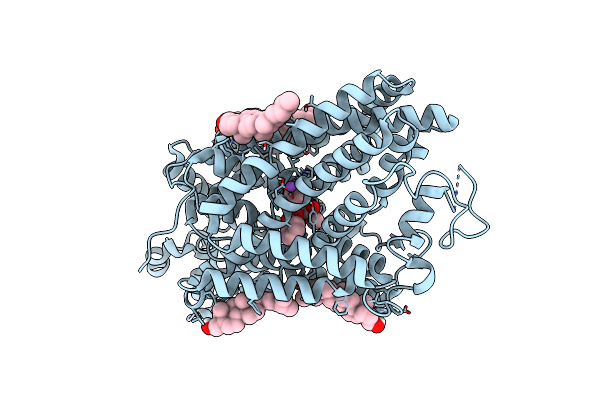 |
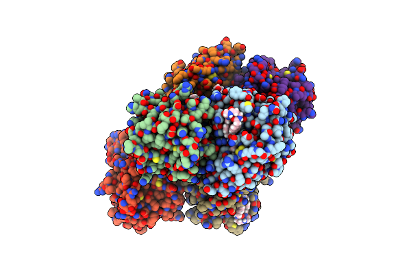
Organism: Homo sapiens
Method: ELECTRON MICROSCOPY Release Date: 2024-07-03 Classification: MEMBRANE PROTEIN Ligands: CLR, Y01, NA, CL, COC |
 |
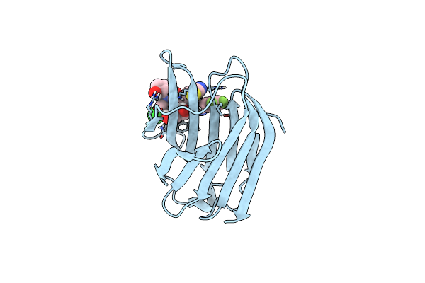
Organism: Severe acute respiratory syndrome coronavirus 2
Method: X-RAY DIFFRACTION Resolution:1.66 Å Release Date: 2024-02-14 Classification: VIRAL PROTEIN Ligands: XWH, DMS, NA, CL |
 |
Organism: Severe acute respiratory syndrome coronavirus 2
Method: X-RAY DIFFRACTION Resolution:1.84 Å Release Date: 2024-02-14 Classification: VIRAL PROTEIN Ligands: XWZ, DMS, CL, NA |
 |
Organism: Severe acute respiratory syndrome coronavirus 2
Method: X-RAY DIFFRACTION Resolution:1.54 Å Release Date: 2024-02-14 Classification: VIRAL PROTEIN Ligands: Y2C, DMS, NA, CL |
 |
Organism: Severe acute respiratory syndrome coronavirus 2
Method: X-RAY DIFFRACTION Resolution:1.87 Å Release Date: 2024-02-14 Classification: VIRAL PROTEIN Ligands: DMS, Y25, NA |
 |
Organism: Severe acute respiratory syndrome coronavirus 2
Method: X-RAY DIFFRACTION Resolution:1.79 Å Release Date: 2024-02-14 Classification: VIRAL PROTEIN Ligands: DMS, Y1R, NA |
 |
Organism: Severe acute respiratory syndrome coronavirus 2
Method: X-RAY DIFFRACTION Resolution:1.74 Å Release Date: 2024-02-14 Classification: VIRAL PROTEIN Ligands: DMS, Y1L, NA |
 |
Organism: Severe acute respiratory syndrome coronavirus 2
Method: X-RAY DIFFRACTION Resolution:1.80 Å Release Date: 2024-02-14 Classification: VIRAL PROTEIN Ligands: DMS, Y1H, CL, NA |
 |
Organism: Severe acute respiratory syndrome coronavirus 2
Method: X-RAY DIFFRACTION Resolution:1.68 Å Release Date: 2024-02-14 Classification: VIRAL PROTEIN Ligands: Y1C, DMS, CL, NA |