
Search Count: 32
 |
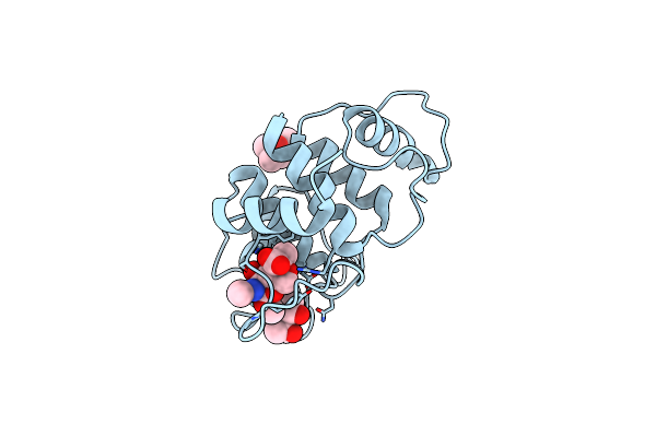
Organism: Paenibacillus alvei
Method: X-RAY DIFFRACTION Resolution:1.90 Å Release Date: 2018-08-15 Classification: SUGAR BINDING PROTEIN Ligands: SO4, CL |
 |
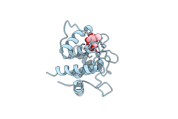
Organism: Paenibacillus alvei
Method: X-RAY DIFFRACTION Resolution:2.25 Å Release Date: 2018-08-15 Classification: SUGAR BINDING PROTEIN Ligands: 6LA |
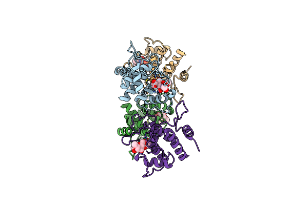 |
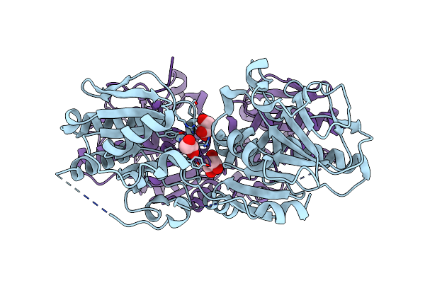
Crystal Structure Of Spaa-Slh In Complex With 4,6-Pyr-Beta-D-Mannacome (P1)
Organism: Paenibacillus alvei
Method: X-RAY DIFFRACTION Resolution:2.00 Å Release Date: 2018-08-15 Classification: SUGAR BINDING PROTEIN Ligands: 6LA |
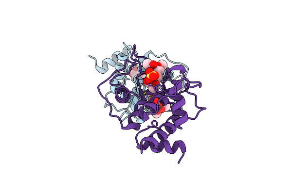 |
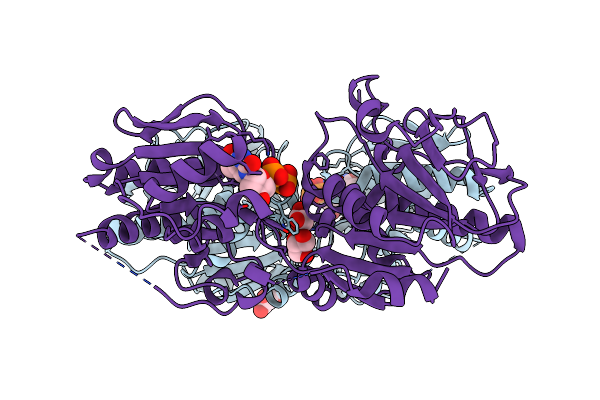
Crystal Structure Of Spaa-Slh In Complex With 4,6-Pyr-Beta-D-Mannacome (C2)
Organism: Paenibacillus alvei
Method: X-RAY DIFFRACTION Resolution:2.16 Å Release Date: 2018-08-15 Classification: SUGAR BINDING PROTEIN Ligands: 6LA, SO4 |
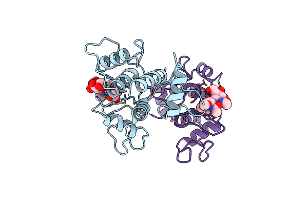 |
Crystal Structure Of Spaa-Slh In Complex With Beta-D-Glcnac-(1->3)-4,6-Pyr-Beta-D-Mannacome
Organism: Paenibacillus alvei
Method: X-RAY DIFFRACTION Resolution:2.15 Å Release Date: 2018-08-15 Classification: SUGAR BINDING PROTEIN Ligands: FHY |
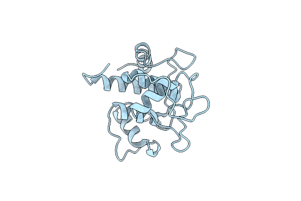 |
Organism: Paenibacillus alvei
Method: X-RAY DIFFRACTION Resolution:1.15 Å Release Date: 2018-08-15 Classification: SUGAR BINDING PROTEIN Ligands: CL |
 |
Crystal Structure Of Spaa-Slh/G109A In Complex With 4,6-Pyr-Beta-D-Mannacome
Organism: Paenibacillus alvei
Method: X-RAY DIFFRACTION Resolution:1.53 Å Release Date: 2018-08-15 Classification: SUGAR BINDING PROTEIN Ligands: 6LA, MPD |
 |
Crystal Structure Of Spaa-Slh/G46A/G109A In Complex With 4,6-Pyr-Beta-D-Mannacome
Organism: Paenibacillus alvei
Method: X-RAY DIFFRACTION Resolution:1.24 Å Release Date: 2018-08-15 Classification: SUGAR BINDING PROTEIN Ligands: 6LA |
 |
Organism: Geobacillus stearothermophilus
Method: X-RAY DIFFRACTION Resolution:2.28 Å Release Date: 2010-02-02 Classification: TRANSFERASE Ligands: GOL |
 |
Organism: Geobacillus stearothermophilus
Method: X-RAY DIFFRACTION Resolution:2.81 Å Release Date: 2010-02-02 Classification: TRANSFERASE Ligands: TYD, GOL |
 |
Organism: Geobacillus stearothermophilus
Method: X-RAY DIFFRACTION Resolution:2.55 Å Release Date: 2010-02-02 Classification: TRANSFERASE Ligands: GOL, TRH |
 |
X-Ray Structure Of Qdtb From T. Thermosaccharolyticum In Complex With A Plp:Tdp-3-Aminoquinovose Aldimine
Organism: Thermoanaerobacterium thermosaccharolyticum
Method: X-RAY DIFFRACTION Resolution:2.15 Å Release Date: 2009-02-17 Classification: TRANSFERASE Ligands: TQP |
 |
Crystal Structure Of Qdtc, The Dtdp-3-Amino-3,6-Dideoxy-D-Glucose N-Acetyl Transferase From Thermoanaerobacterium Thermosaccharolyticum In Complex With Acetyl-Coa
Organism: Thermoanaerobacterium thermosaccharolyticum
Method: X-RAY DIFFRACTION Resolution:1.70 Å Release Date: 2009-02-17 Classification: TRANSFERASE Ligands: ACO, TDR |
 |
Crystal Structure Of Qdtc, The Dtdp-3-Amino-3,6-Dideoxy-D-Glucose N-Acetyl Transferase From Thermoanaerobacterium Thermosaccharolyticum In Complex With Coa And Dtdp-3-Amino-Quinovose
Organism: Thermoanaerobacterium thermosaccharolyticum
Method: X-RAY DIFFRACTION Resolution:1.95 Å Release Date: 2009-02-17 Classification: TRANSFERASE Ligands: COA, T3Q |
 |
Crystal Structure Of Qdtc, The Dtdp-3-Amino-3,6-Dideoxy-D-Glucose N-Acetyl Transferase From Thermoanaerobacterium Thermosaccharolyticum In Complex With Coa And Dtdp-3-Amino-Fucose
Organism: Thermoanaerobacterium thermosaccharolyticum
Method: X-RAY DIFFRACTION Resolution:1.80 Å Release Date: 2009-02-17 Classification: TRANSFERASE Ligands: COA, T3F |
 |
Organism: Aneurinibacillus thermoaerophilus
Method: X-RAY DIFFRACTION Resolution:1.82 Å Release Date: 2008-03-25 Classification: OXIDOREDUCTASE Ligands: A2R, GDD |
 |
Organism: Mycobacterium tuberculosis
Method: X-RAY DIFFRACTION Resolution:1.79 Å Release Date: 2006-10-26 Classification: ISOMERASE Ligands: TRH |
 |
Organism: Pseudomonas aeruginosa
Method: X-RAY DIFFRACTION Resolution:2.00 Å Release Date: 2006-10-26 Classification: ISOMERASE Ligands: TRH |
 |
Organism: Streptococcus suis
Method: X-RAY DIFFRACTION Resolution:1.60 Å Release Date: 2006-10-26 Classification: ISOMERASE Ligands: TRH, NI |
 |
Organism: Pseudomonas aeruginosa
Method: X-RAY DIFFRACTION Release Date: 2006-08-14 Classification: ISOMERASE Ligands: TDO |