
Search Count: 15
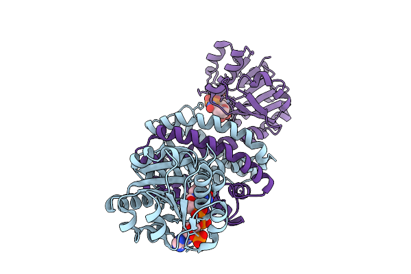 |
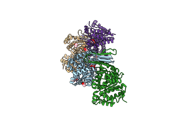
Organism: Kutzneria albida dsm 43870
Method: X-RAY DIFFRACTION Release Date: 2025-07-30 Classification: OXIDOREDUCTASE Ligands: NDP, NA |
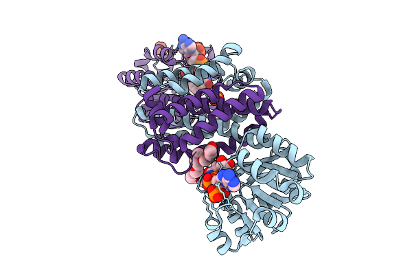 |
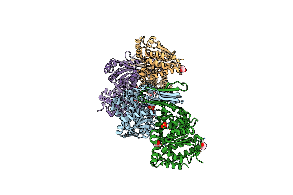
Organism: Kutzneria albida dsm 43870
Method: X-RAY DIFFRACTION Release Date: 2025-07-30 Classification: OXIDOREDUCTASE Ligands: NDP, SO4, PE8, NA |
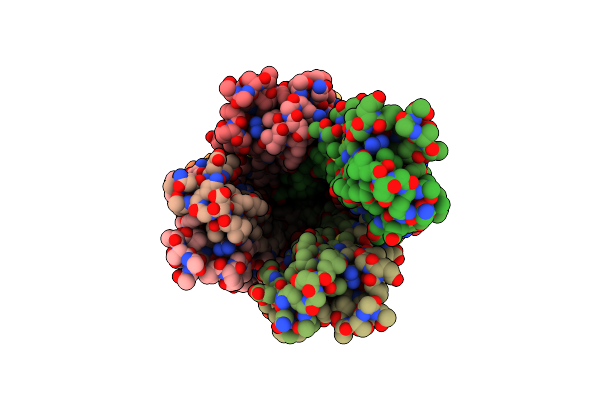 |
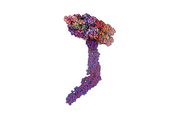
Organism: Acinetobacter genomosp. 16bj
Method: ELECTRON MICROSCOPY Release Date: 2024-03-06 Classification: CELL ADHESION |
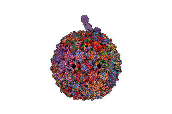 |
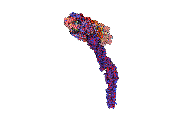
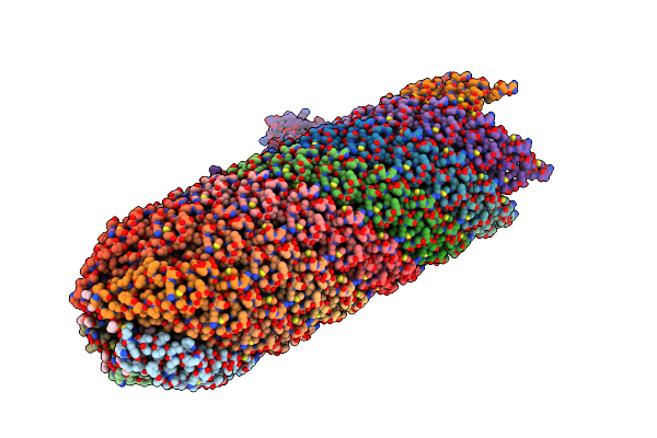
Organism: Bacteria abnormis, Acinetobacter phage ap205
Method: ELECTRON MICROSCOPY Release Date: 2024-03-06 Classification: VIRUS/RNA |
 |
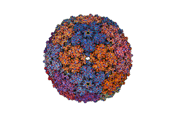
Organism: Acinetobacter phage ap205, Acinetobacter genomosp. 16bj
Method: ELECTRON MICROSCOPY Release Date: 2024-03-06 Classification: VIRUS |
 |
Organism: Acinetobacter phage ap205, Acinetobacter genomosp. 16bj
Method: ELECTRON MICROSCOPY Release Date: 2024-03-06 Classification: VIRUS |
 |
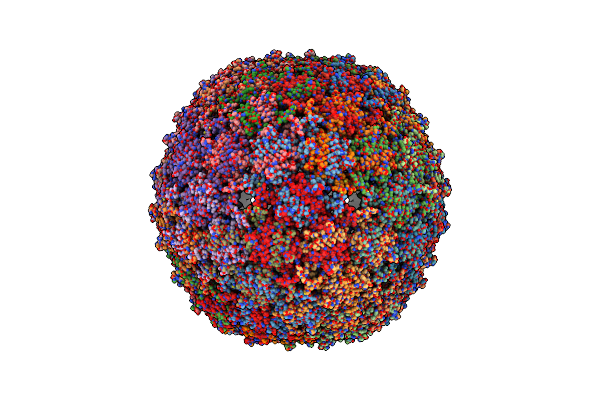
Organism: Acinetobacter phage ap205
Method: ELECTRON MICROSCOPY Release Date: 2024-03-06 Classification: VIRUS LIKE PARTICLE |
 |
Organism: Acinetobacter phage ap205
Method: ELECTRON MICROSCOPY Release Date: 2024-03-06 Classification: VIRUS LIKE PARTICLE |
 |
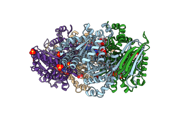
Crystal Structure Of Isocitrate Dehydrogenase From Ostreococcus Tauri In Complex With Isocitrate And Magnesium(Ii)
Organism: Ostreococcus tauri
Method: X-RAY DIFFRACTION Resolution:1.80 Å Release Date: 2021-05-19 Classification: OXIDOREDUCTASE Ligands: ICT, MG, FLC, IPA, GOL |
 |
Organism: Ostreococcus tauri
Method: X-RAY DIFFRACTION Resolution:1.75 Å Release Date: 2019-12-18 Classification: OXIDOREDUCTASE Ligands: GOL, SO4 |
 |
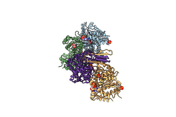
Crystal Structure Of Isocitrate Dehydrogenase From Ostreococcus Tauri In Complex With Nad+ And Citrate
Organism: Ostreococcus tauri
Method: X-RAY DIFFRACTION Resolution:1.87 Å Release Date: 2019-12-18 Classification: OXIDOREDUCTASE Ligands: SO4, GOL, FLC, NAD, PEG |
 |
Crystal Structure Of Isocitrate Dehydrogenase From Ostreococcus Tauri In Complex With Nad+ And Mg2+
Organism: Ostreococcus tauri
Method: X-RAY DIFFRACTION Resolution:1.78 Å Release Date: 2019-12-18 Classification: OXIDOREDUCTASE Ligands: NAD, MG, GOL, SO4 |
 |
Organism: Escherichia coli, Enterobacteria phage ms2
Method: ELECTRON MICROSCOPY Release Date: 2019-07-24 Classification: PROTEIN BINDING Ligands: KSV |
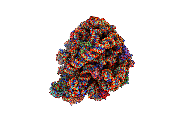 |
Cryo-Em Structure Of The Large Ribosomal Subunit From Mycobacterium Tuberculosis Bound With A Potent Linezolid Analog
Organism: Mycobacterium tuberculosis
Method: ELECTRON MICROSCOPY Release Date: 2017-09-20 Classification: RIBOSOME Ligands: 917 |
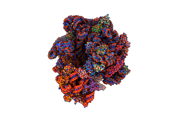 |
Cryo-Em Structure Of The 70S Ribosome From Mycobacterium Tuberculosis Bound With Capreomycin
Organism: Mycobacterium tuberculosis, Streptomyces puniceus
Method: ELECTRON MICROSCOPY Release Date: 2017-09-20 Classification: RIBOSOME |