
Search Count: 13
 |
Organism: Alicyclobacillus acidocaldarius
Method: X-RAY DIFFRACTION Resolution:2.00 Å Release Date: 2007-06-26 Classification: HYDROLASE Ligands: SO4 |
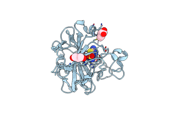 |
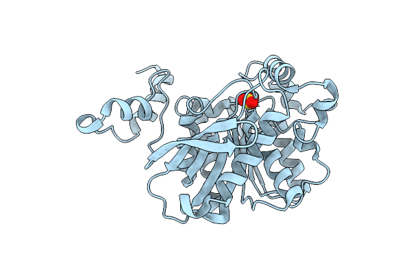
Crystal Structure Of The Human Carbonic Anhydrase Ii In Complex With The 5-Amino-1,3,4-Thiadiazole-2-Sulfonamide Inhibitor.
Organism: Homo sapiens
Method: X-RAY DIFFRACTION Resolution:1.55 Å Release Date: 2006-12-19 Classification: LYASE Ligands: ZN, 1SA, MBO, GOL |
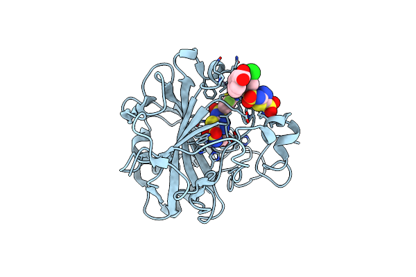 |
Crystal Structure Of The Human Carbonic Anhydrase Ii In Complex With The 5-(4-Amino-3-Chloro-5-Fluorophenylsulfonamido)-1,3,4-Thiadiazole-2-Sulfonamide Inhibitor
Organism: Homo sapiens
Method: X-RAY DIFFRACTION Resolution:2.10 Å Release Date: 2006-10-10 Classification: LYASE Ligands: ZN, 1CN, MBO, GOL |
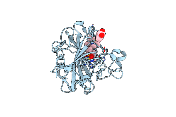 |
Carbonic Anhydrase Ii In Complex With A Membrane-Impermeant Sulfonamide Inhibitor
Organism: Homo sapiens
Method: X-RAY DIFFRACTION Resolution:2.00 Å Release Date: 2005-10-18 Classification: LYASE Ligands: ZN, HGB, PIU, GOL |
 |
Organism: Alicyclobacillus acidocaldarius
Method: X-RAY DIFFRACTION Resolution:2.10 Å Release Date: 2004-10-05 Classification: HYDROLASE Ligands: SO4 |
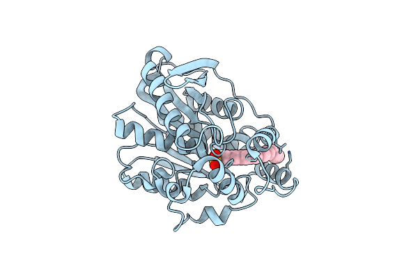 |
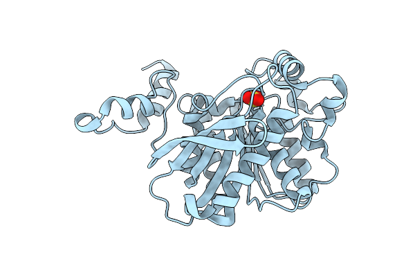
Crystal Structure Of Mutant M211S/R215L Of Carboxylesterase Est2 Complexed With Hexadecanesulfonate
Organism: Alicyclobacillus acidocaldarius
Method: X-RAY DIFFRACTION Resolution:2.30 Å Release Date: 2004-03-23 Classification: HYDROLASE Ligands: HDS |
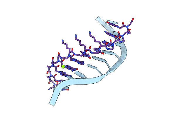 |
Method: X-RAY DIFFRACTION
Release Date: 2003-10-28 Classification: DNA/PEPTIDE NUCLEIC ACID Ligands: MG |
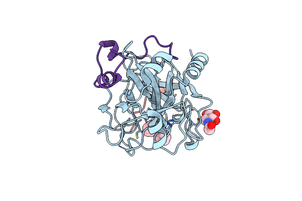 |
Design Of Weakly Basic Thrombin Inhibitors Incorporating Novel P1 Binding Functions: Molecular And X-Ray Crystallographic Studies.
Organism: Hirudo medicinalis, Homo sapiens
Method: X-RAY DIFFRACTION Resolution:1.90 Å Release Date: 2003-08-26 Classification: HYDROLASE/HYDROLASE INHIBITOR Ligands: NAG, 4ND |
 |
The Crystal Structure Of The K18G Mutant Of The Thioredoxin From Alicyclobacillus Acidocaldarius
Organism: Alicyclobacillus acidocaldarius
Method: X-RAY DIFFRACTION Resolution:1.90 Å Release Date: 2003-08-05 Classification: ELECTRON TRANSPORT |
 |
The Crystal Structure Of The Mutant R82E Of Thioredoxin From Alicyclobacillus Acidocaldarius
Organism: Alicyclobacillus acidocaldarius
Method: X-RAY DIFFRACTION Resolution:1.90 Å Release Date: 2003-08-05 Classification: ELECTRON TRANSPORT Ligands: ZN, ACT, CAC |
 |
Organism: Chlamydomonas reinhardtii
Method: X-RAY DIFFRACTION Resolution:2.10 Å Release Date: 2001-12-12 Classification: ELECTRON TRANSPORT |
 |
Crystal Structure Of A Mutated Thioredoxin, D30A, From Chlamydomonas Reinhardtii
Organism: Chlamydomonas reinhardtii
Method: X-RAY DIFFRACTION Resolution:2.20 Å Release Date: 2001-12-12 Classification: ELECTRON TRANSPORT |
 |
The Crystal Structure Of A Hyper-Thermophilic Carboxylesterase From The Archaeon Archaeoglobus Fulgidus
Organism: Archaeoglobus fulgidus
Method: X-RAY DIFFRACTION Resolution:2.20 Å Release Date: 2001-12-05 Classification: HYDROLASE Ligands: EPE |