
Search Count: 18
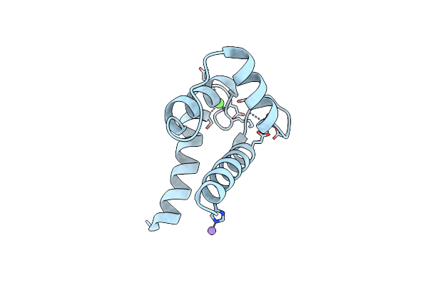 |
Organism: Bos taurus
Method: X-RAY DIFFRACTION Resolution:2.17 Å Release Date: 2024-02-07 Classification: METAL BINDING PROTEIN Ligands: CA, MN |
 |
Organism: Homo sapiens
Method: ELECTRON MICROSCOPY Release Date: 2022-08-03 Classification: MEMBRANE PROTEIN Ligands: ZN, ATP |
 |
Organism: Homo sapiens
Method: ELECTRON MICROSCOPY Release Date: 2022-08-03 Classification: MEMBRANE PROTEIN Ligands: ZN, ATP, CA, XAN |
 |
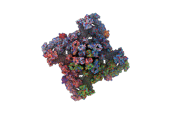
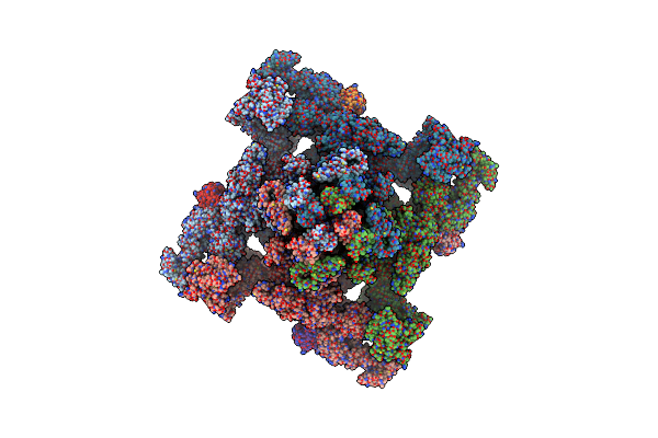
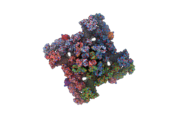
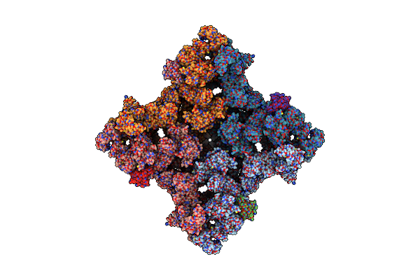
Structure Of Pka Phosphorylated Human Ryr2 In The Closed State In The Presence Of Calmodulin
Organism: Homo sapiens
Method: ELECTRON MICROSCOPY Release Date: 2022-08-03 Classification: MEMBRANE PROTEIN Ligands: ZN, ATP |
 |
Organism: Homo sapiens
Method: ELECTRON MICROSCOPY Release Date: 2022-08-03 Classification: MEMBRANE PROTEIN Ligands: ZN, ATP |
 |
Organism: Homo sapiens
Method: ELECTRON MICROSCOPY Release Date: 2022-08-03 Classification: MEMBRANE PROTEIN Ligands: ZN, ATP, CA, XAN |
 |
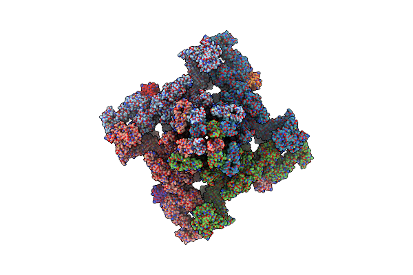
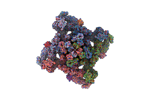
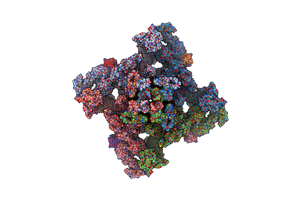
Structure Of Pka Phosphorylated Human Ryr2-R2474S In The Closed State In The Presence Of Arm210
Organism: Homo sapiens
Method: ELECTRON MICROSCOPY Release Date: 2022-08-03 Classification: MEMBRANE PROTEIN Ligands: ZN, ATP, KVR |
 |
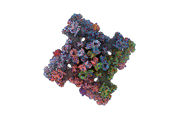
Structure Of Pka Phosphorylated Human Ryr2-R2474S In The Closed State In The Presence Of Calmodulin
Organism: Homo sapiens
Method: ELECTRON MICROSCOPY Release Date: 2022-08-03 Classification: MEMBRANE PROTEIN Ligands: ZN, ATP |
 |
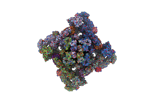
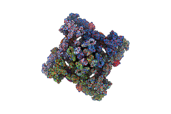
Structure Of Pka Phosphorylated Human Ryr2-R2474S In The Open State In The Presence Of Calmodulin
Organism: Homo sapiens
Method: ELECTRON MICROSCOPY Release Date: 2022-08-03 Classification: MEMBRANE PROTEIN Ligands: ZN, ATP, CA, XAN |
 |
Organism: Homo sapiens
Method: ELECTRON MICROSCOPY Release Date: 2022-08-03 Classification: MEMBRANE PROTEIN Ligands: ZN, ATP |
 |
Organism: Homo sapiens
Method: ELECTRON MICROSCOPY Release Date: 2022-08-03 Classification: MEMBRANE PROTEIN Ligands: ZN, ATP, CA, XAN |
 |
Organism: Homo sapiens, Oryctolagus cuniculus
Method: ELECTRON MICROSCOPY Release Date: 2022-05-18 Classification: MEMBRANE PROTEIN Ligands: CA, ATP, ZN, CFF, KVR, L9R |
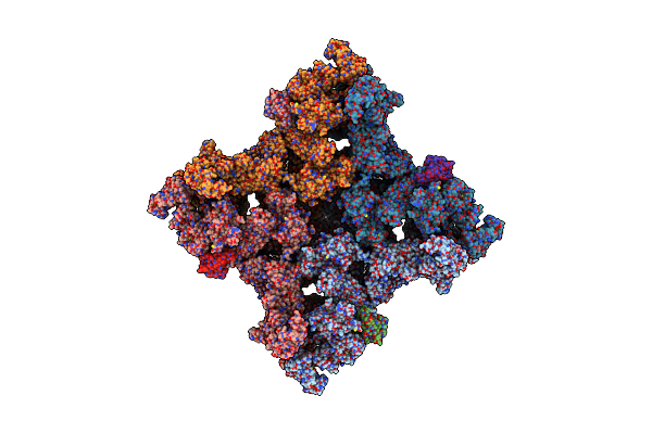 |
High Resolution Structure Of The Membrane Embedded Skeletal Muscle Ryanodine Receptor
Organism: Homo sapiens, Oryctolagus cuniculus
Method: ELECTRON MICROSCOPY Release Date: 2021-09-08 Classification: MEMBRANE PROTEIN/ISOMERASE Ligands: ATP, CA, ZN, CFF |
 |
High Resolution Structure Of The Membrane Embedded Skeletal Muscle Ryanodine Receptor
Organism: Homo sapiens, Oryctolagus cuniculus
Method: ELECTRON MICROSCOPY Release Date: 2021-08-18 Classification: MEMBRANE PROTEIN/ISOMERASE Ligands: ATP, CA, ZN, CFF |
 |
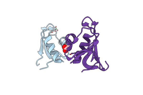
Organism: Homo sapiens
Method: X-RAY DIFFRACTION Resolution:2.25 Å Release Date: 2017-04-12 Classification: METAL BINDING PROTEIN Ligands: TRS, CA |
 |
Organism: Homo sapiens
Method: X-RAY DIFFRACTION Resolution:1.77 Å Release Date: 2017-04-12 Classification: RNA BINDING PROTEIN Ligands: ACT, NI |
 |
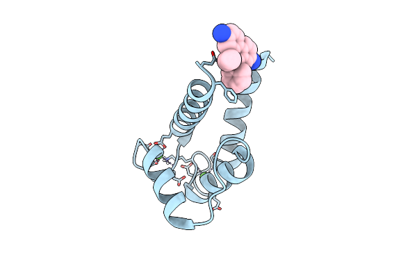
Organism: Bos taurus
Method: X-RAY DIFFRACTION Resolution:1.81 Å Release Date: 2016-06-08 Classification: METAL BINDING PROTEIN/INHIBITOR Ligands: CA, 5RL |
 |
Organism: Bos taurus
Method: X-RAY DIFFRACTION Resolution:1.26 Å Release Date: 2016-06-08 Classification: METAL BINDING PROTEIN/INHIBITOR Ligands: CA, ET |