
Search Count: 13
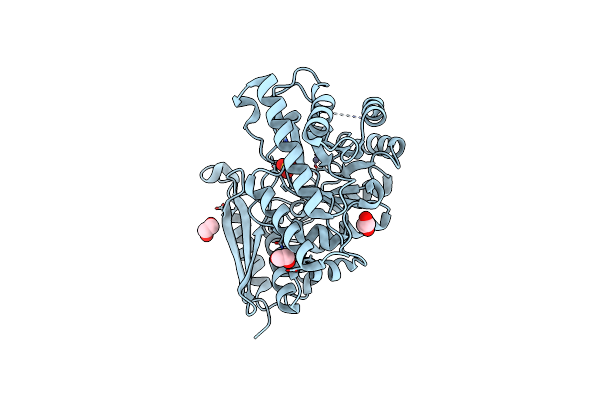 |
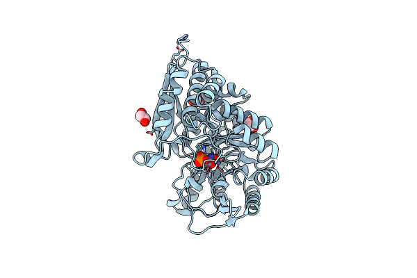
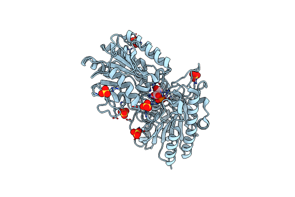
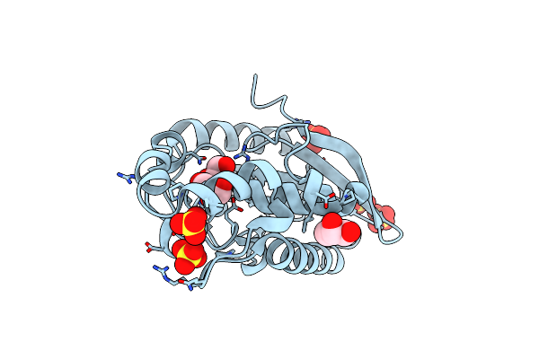
Crystal Structure Of The T. Brucei Enolase Complexed With Sulphate, Identification Of A Metal Binding Site Iv
Organism: Trypanosoma brucei
Method: X-RAY DIFFRACTION Resolution:1.90 Å Release Date: 2007-11-20 Classification: LYASE Ligands: ZN, SO4, EDO |
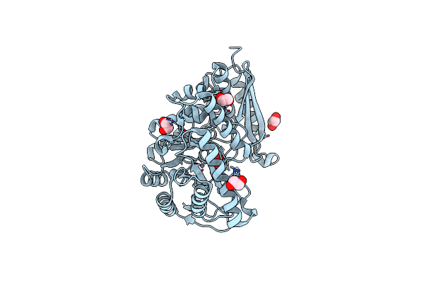 |
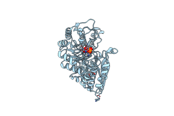
Crystal Structure Of The T. Brucei Enolase Complexed With Sulphate In Closed Conformation
Organism: Trypanosoma brucei
Method: X-RAY DIFFRACTION Resolution:1.90 Å Release Date: 2007-11-20 Classification: LYASE Ligands: ZN, SO4, EDO |
 |
Organism: Trypanosoma brucei
Method: X-RAY DIFFRACTION Resolution:2.00 Å Release Date: 2007-11-20 Classification: LYASE Ligands: ZN, PEP, EDO |
 |
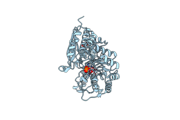
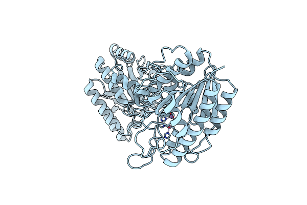
Crystal Structure Of The T. Brucei Enolase Complexed With Phosphonoacetohydroxamate (Pah), His156-Out Conformation
Organism: Trypanosoma brucei
Method: X-RAY DIFFRACTION Resolution:1.65 Å Release Date: 2007-11-20 Classification: LYASE Ligands: ZN, PAH, EDO |
 |
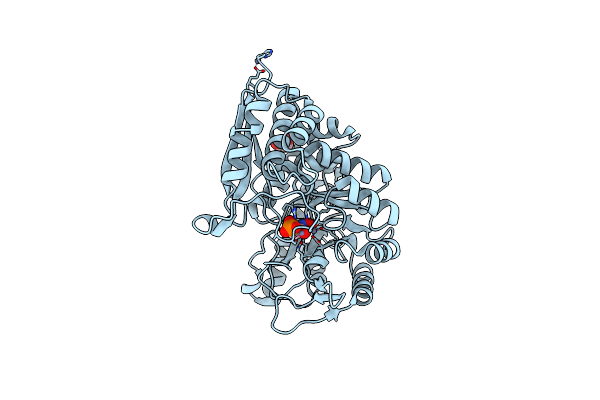
Crystal Structure Of The T. Brucei Enolase Complexed With Phosphonoacetohydroxamate (Pah), His156-In Conformation
Organism: Trypanosoma brucei
Method: X-RAY DIFFRACTION Resolution:1.90 Å Release Date: 2007-11-20 Classification: LYASE Ligands: ZN, PAH, EDO |
 |
Crystal Structure Of The T. Brucei Enolase Complexed With Fluoro-Phosphonoacetohydroxamate (Fpah)
Organism: Trypanosoma brucei
Method: X-RAY DIFFRACTION Resolution:1.80 Å Release Date: 2007-11-20 Classification: LYASE Ligands: ZN, FSG, EDO |
 |
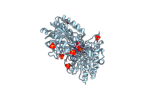
Structure Of Bacillus Anthracis Cofactor-Independent Phosphoglucerate Mutase
Organism: Bacillus anthracis
Method: X-RAY DIFFRACTION Resolution:2.38 Å Release Date: 2006-10-24 Classification: ISOMERASE Ligands: MN |
 |
1.4A Crystal Structure Of Phosphoglycerate Mutase From Bacillus Stearothermophilus Complexed With 2-Phosphoglycerate
Organism: Bacillus stearothermophilus
Method: X-RAY DIFFRACTION Resolution:1.40 Å Release Date: 2003-05-01 Classification: ISOMERASE Ligands: MN, 2PG, SO4 |
 |
Crystal Structure Of The S62A Mutant Of Phosphoglycerate Mutase From Bacillus Stearothermophilus Complexed With 2-Phosphoglycerate
Organism: Bacillus stearothermophilus
Method: X-RAY DIFFRACTION Resolution:2.65 Å Release Date: 2002-12-23 Classification: ISOMERASE Ligands: MN, 2PG, SO4 |
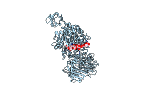 |
Crystal Structure Of Streptococcus Agalactiae Hyaluronate Lyase Complexed With Hexasaccharide Unit Of Hyaluronan
Organism: Streptococcus agalactiae
Method: X-RAY DIFFRACTION Resolution:2.20 Å Release Date: 2002-10-30 Classification: LYASE |
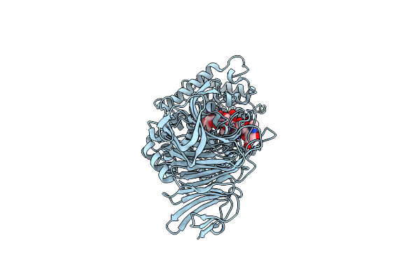 |
Streptococcus Pneumoniae Hyaluronate Lyase In Complex With Hexasaccharide Hyaluronan Substrate
Organism: Streptococcus pneumoniae
Method: X-RAY DIFFRACTION Resolution:2.00 Å Release Date: 2002-08-07 Classification: LYASE |
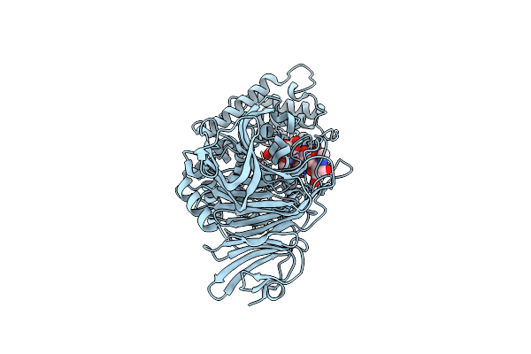 |
Streptococcus Pneumoniae Hyaluronate Lyase In Complex With Tetrasaccharide Hyaluronan Substrate
Organism: Streptococcus pneumoniae
Method: X-RAY DIFFRACTION Resolution:1.53 Å Release Date: 2002-08-07 Classification: LYASE |
 |
Organism: Bacillus stearothermophilus
Method: X-RAY DIFFRACTION Resolution:2.30 Å Release Date: 2002-02-11 Classification: HYDROLASE Ligands: SO4, GOL |