
Search Count: 22
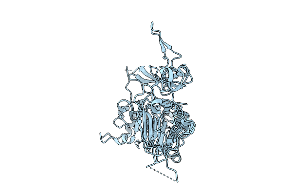 |
Her2/Erbb2 Extracellular Domain (Ecd) From A Near Full-Length Construct Solubilized In Amphipols.
Organism: Homo sapiens, Aequorea victoria
Method: ELECTRON MICROSCOPY Release Date: 2025-08-13 Classification: SIGNALING PROTEIN |
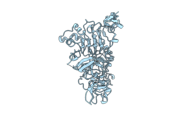 |
Her2/Erbb2 Extracellular Domain (Ecd) In Compact Conformation In Complex With Trastuzumab (Tzb) Antibody
Organism: Homo sapiens, Aequorea victoria
Method: ELECTRON MICROSCOPY Release Date: 2025-08-06 Classification: SIGNALING PROTEIN |
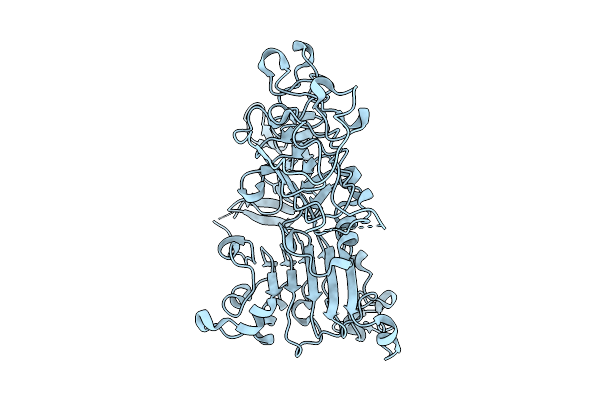 |
Her2/Erbb2 Extracellular Domain (Ecd) In Extended Conformation In Complex With Trastuzumab (Tzb) Antibody
Organism: Homo sapiens, Aequorea victoria
Method: ELECTRON MICROSCOPY Release Date: 2025-08-06 Classification: SIGNALING PROTEIN |
 |
Cryo-Em Structure Of Brucella Abortus Lumazine Synthase (Bls) Engineered With Shiga Toxin I Subunit B (Stx1B)
Organism: Escherichia coli o157:h7, Brucella abortus biovar 1 (strain 9-941)
Method: ELECTRON MICROSCOPY Release Date: 2025-03-26 Classification: TOXIN |
 |
Cryo-Em Structure Of Brucella Abortus Lumazine Synthase (Bls) Engineered With Shiga Toxin Ii Subunit B (Stx2B)
Organism: Escherichia coli o157:h7, Brucella abortus bv. 1 str. 9-941
Method: ELECTRON MICROSCOPY Release Date: 2025-03-26 Classification: TOXIN |
 |
Organism: Trypanosoma brucei brucei
Method: ELECTRON MICROSCOPY Release Date: 2024-10-02 Classification: MEMBRANE PROTEIN Ligands: VO6 |
 |
Organism: Trypanosoma brucei brucei
Method: ELECTRON MICROSCOPY Release Date: 2024-10-02 Classification: MEMBRANE PROTEIN Ligands: PNT |
 |
Organism: Trypanosoma brucei brucei
Method: ELECTRON MICROSCOPY Release Date: 2024-10-02 Classification: MEMBRANE PROTEIN Ligands: GOL |
 |
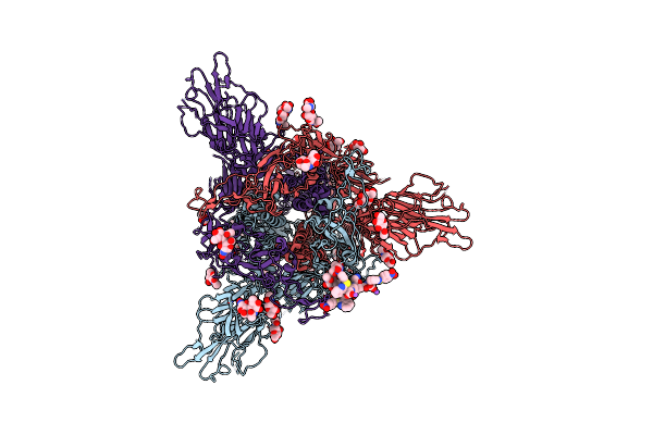
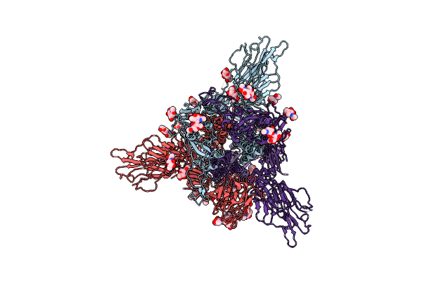
Organism: Severe acute respiratory syndrome coronavirus 2
Method: ELECTRON MICROSCOPY Resolution:3.40 Å Release Date: 2023-09-27 Classification: VIRAL PROTEIN Ligands: NAG |
 |
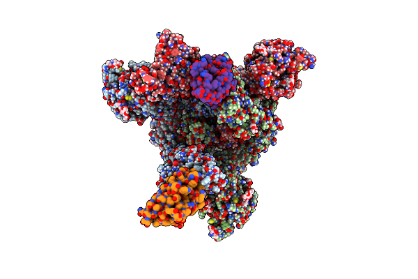
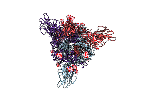
Sars-Cov-2 S Protein S:D614G Mutant In 3-Down With Binding Site Of An Entry Inhibitor
Organism: Severe acute respiratory syndrome coronavirus 2
Method: ELECTRON MICROSCOPY Resolution:4.30 Å Release Date: 2023-09-27 Classification: VIRAL PROTEIN Ligands: NAG, NA, XIO |
 |
Cryo-Em Structure Of Mal De Rio Cuarto Virus P9-1 Viroplasm Protein (Decamer)
Organism: Mal de rio cuarto virus
Method: ELECTRON MICROSCOPY Release Date: 2022-06-15 Classification: VIRAL PROTEIN |
 |
Cryo-Em Structure Of Mal De Rio Cuarto Virus P9-1 Viroplasm Protein (Dodecamer)
Organism: Mal de rio cuarto virus
Method: ELECTRON MICROSCOPY Release Date: 2022-06-15 Classification: VIRAL PROTEIN |
 |
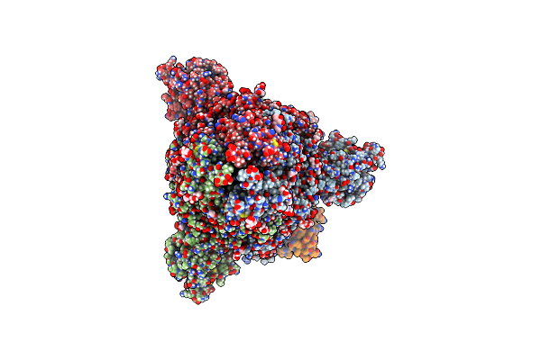
Organism: Severe acute respiratory syndrome coronavirus 2, Camelus dromedarius
Method: ELECTRON MICROSCOPY Release Date: 2022-06-08 Classification: VIRAL PROTEIN Ligands: NAG |
 |
Organism: Severe acute respiratory syndrome coronavirus 2, Camelus dromedarius
Method: ELECTRON MICROSCOPY Release Date: 2022-06-08 Classification: VIRAL PROTEIN Ligands: NAG |
 |
Organism: Severe acute respiratory syndrome coronavirus 2, Camelus dromedarius
Method: ELECTRON MICROSCOPY Release Date: 2022-06-08 Classification: VIRAL PROTEIN Ligands: NAG |
 |
Organism: Severe acute respiratory syndrome coronavirus 2, Escherichia virus t4
Method: ELECTRON MICROSCOPY Resolution:3.40 Å Release Date: 2022-05-25 Classification: VIRAL PROTEIN Ligands: NAG |
 |
Organism: Severe acute respiratory syndrome coronavirus 2, Escherichia virus t4
Method: ELECTRON MICROSCOPY Resolution:4.20 Å Release Date: 2022-05-25 Classification: VIRAL PROTEIN Ligands: NAG |
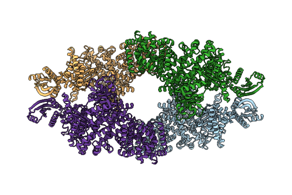 |
Cryo-Em Map Of The Large Glutamate Dehydrogenase Composed Of 180 Kda Subunits From Mycobacterium Smegmatis (Open Conformation)
Organism: Mycolicibacterium smegmatis mc2 155
Method: ELECTRON MICROSCOPY Release Date: 2021-06-09 Classification: OXIDOREDUCTASE |
 |
Crystal Structure Of The Large Glutamate Dehydrogenase Composed Of 180 Kda Subunits From Mycobacterium Smegmatis
Organism: Mycolicibacterium smegmatis
Method: X-RAY DIFFRACTION Resolution:6.27 Å Release Date: 2021-06-09 Classification: OXIDOREDUCTASE |
 |
Organism: Severe acute respiratory syndrome coronavirus 2
Method: ELECTRON MICROSCOPY Release Date: 2020-07-29 Classification: VIRAL PROTEIN Ligands: NAG, MAN, DMS |