
Search Count: 28
 |
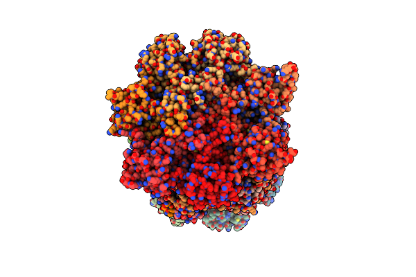
Organism: Spinacia oleracea
Method: ELECTRON MICROSCOPY Release Date: 2022-08-24 Classification: PLANT PROTEIN Ligands: ATP, ADP |
 |
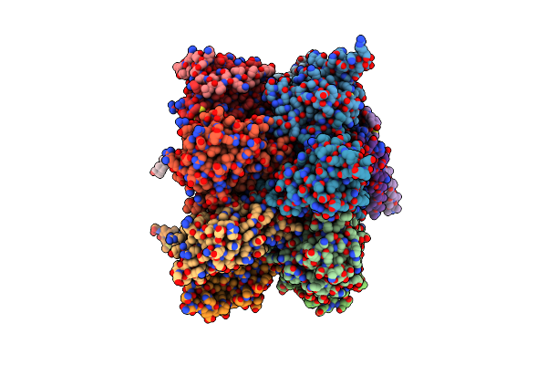
Organism: Xenopus laevis
Method: ELECTRON MICROSCOPY Release Date: 2022-07-20 Classification: RIBOSOME Ligands: MG, ZN |
 |
Organism: Danio rerio, Oryctolagus cuniculus
Method: ELECTRON MICROSCOPY Release Date: 2022-07-20 Classification: RIBOSOME Ligands: MG, ZN |
 |
Organism: Danio rerio
Method: ELECTRON MICROSCOPY Release Date: 2022-07-13 Classification: RIBOSOME Ligands: ZN, MG |
 |
Organism: Danio rerio
Method: ELECTRON MICROSCOPY Release Date: 2022-07-13 Classification: RIBOSOME Ligands: MG, ZN |
 |
Organism: Spinacia oleracea
Method: ELECTRON MICROSCOPY Release Date: 2022-05-25 Classification: PLANT PROTEIN |
 |
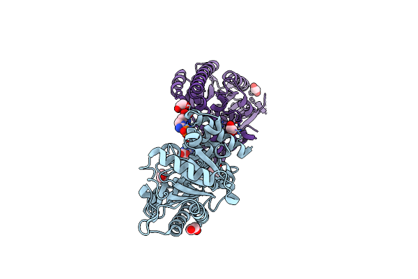
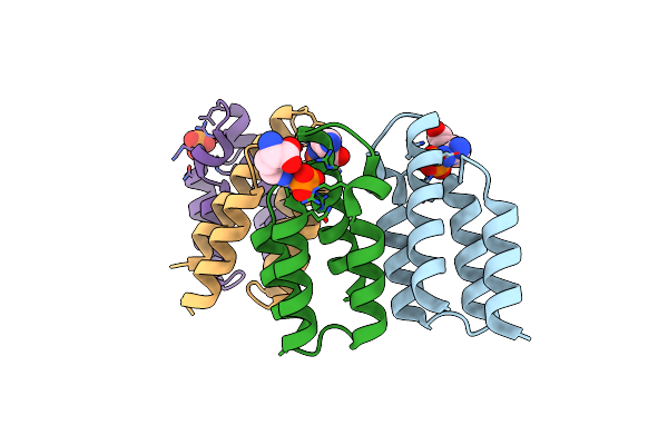
Organism: Homo sapiens
Method: ELECTRON MICROSCOPY Resolution:3.20 Å Release Date: 2021-09-01 Classification: TRANSPORT PROTEIN Ligands: K |
 |
Organism: Drosophila melanogaster
Method: X-RAY DIFFRACTION Resolution:2.50 Å Release Date: 2021-03-03 Classification: MOTOR PROTEIN Ligands: SO4 |
 |
Organism: Drosophila melanogaster
Method: X-RAY DIFFRACTION Resolution:1.42 Å Release Date: 2021-03-03 Classification: MOTOR PROTEIN |
 |
Organism: Drosophila melanogaster
Method: X-RAY DIFFRACTION Resolution:1.79 Å Release Date: 2021-03-03 Classification: MOTOR PROTEIN Ligands: EDO |
 |
Crystal Structure Of The Spoc Domain Of Human Phf3 In Complex With Rna Polymerase Ii Ctd Diheptapeptide Phosphorylated On Ser2
Organism: Homo sapiens
Method: X-RAY DIFFRACTION Resolution:1.93 Å Release Date: 2020-07-15 Classification: PROTEIN BINDING |
 |
Crystal Structure Of The Spoc Domain Of Human Phf3 In Complex With Rna Polymerase Ii Ctd Diheptapeptide Phosphorylated On Ser2Ser7
Organism: Homo sapiens
Method: X-RAY DIFFRACTION Resolution:1.75 Å Release Date: 2020-07-15 Classification: TRANSCRIPTION |
 |
Organism: Homo sapiens
Method: X-RAY DIFFRACTION Resolution:2.59 Å Release Date: 2020-07-15 Classification: TRANSCRIPTION Ligands: GOL |
 |
Crystal Structure Of The Spoc Domain Of Human Phf3 In Complex With Rna Polymerase Ii Ctd Diheptapeptide Phosphorylated On Ser2Ser5
Organism: Homo sapiens
Method: X-RAY DIFFRACTION Resolution:2.85 Å Release Date: 2020-07-15 Classification: TRANSCRIPTION |
 |
Crystal Structure Of Angel2, A 2',3'-Cyclic Phosphatase, In Complex With Adenosine-2',3'-Vanadate
Organism: Homo sapiens
Method: X-RAY DIFFRACTION Resolution:2.10 Å Release Date: 2020-05-20 Classification: RNA BINDING PROTEIN Ligands: KL2, MG, ADN, K |
 |
Organism: Homo sapiens
Method: X-RAY DIFFRACTION Resolution:1.45 Å Release Date: 2020-05-20 Classification: RNA BINDING PROTEIN Ligands: MG, K, GOL |
 |
Organism: Drosophila melanogaster
Method: X-RAY DIFFRACTION Resolution:2.80 Å Release Date: 2019-07-03 Classification: RNA BINDING PROTEIN |
 |
Organism: Drosophila melanogaster
Method: X-RAY DIFFRACTION Resolution:2.00 Å Release Date: 2019-07-03 Classification: RNA BINDING PROTEIN |
 |
Organism: Geobacillus stearothermophilus
Method: X-RAY DIFFRACTION Resolution:1.85 Å Release Date: 2019-02-27 Classification: SIGNALING PROTEIN Ligands: RPI, EDO |
 |
Organism: Bacillus subtilis
Method: X-RAY DIFFRACTION Resolution:2.49 Å Release Date: 2019-02-27 Classification: SIGNALING PROTEIN Ligands: RPI, PO4 |