
Search Count: 88
 |
Organism: Mus musculus, Synthetic construct
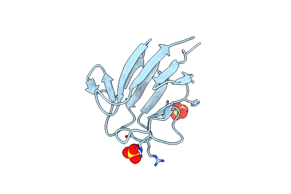
Method: X-RAY DIFFRACTION Resolution:2.35 Å Release Date: 2023-08-30 Classification: DNA BINDING PROTEIN/DNA Ligands: CAC |
 |
Organism: Mus musculus, Synthetic construct
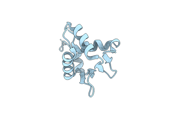
Method: X-RAY DIFFRACTION Resolution:2.30 Å Release Date: 2023-08-30 Classification: DNA BINDING PROTEIN/DNA |
 |
Crystal Structure Of The Helical Cell Shape Determining Protein Pgp2 From Campylobacter Jejuni
Organism: Campylobacter jejuni subsp. jejuni
Method: X-RAY DIFFRACTION Resolution:1.50 Å Release Date: 2021-03-17 Classification: HYDROLASE |
 |
Crystal Structure Of The Helical Cell Shape Determining Protein Pgp2 (K307A Mutant) From Campylobacter Jejuni
Organism: Campylobacter jejuni subsp. jejuni
Method: X-RAY DIFFRACTION Resolution:1.85 Å Release Date: 2021-03-17 Classification: HYDROLASE |
 |
Organism: Homo sapiens
Method: X-RAY DIFFRACTION Resolution:1.85 Å Release Date: 2021-01-20 Classification: ONCOPROTEIN Ligands: FMT |
 |
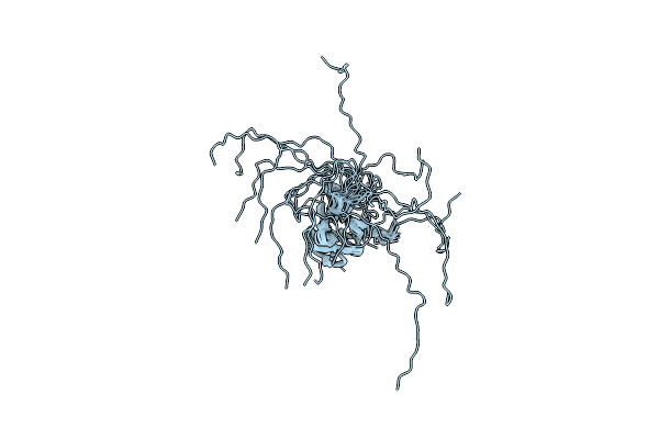
Organism: Lymantria dispar
Method: SOLUTION NMR Release Date: 2020-09-23 Classification: LIPID BINDING PROTEIN |
 |
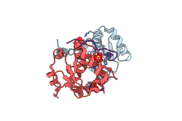
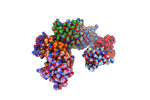
Organism: Salmonella typhimurium (strain lt2 / sgsc1412 / atcc 700720)
Method: X-RAY DIFFRACTION Resolution:1.20 Å Release Date: 2020-09-16 Classification: PROTEIN TRANSPORT Ligands: CD, CL, NA |
 |
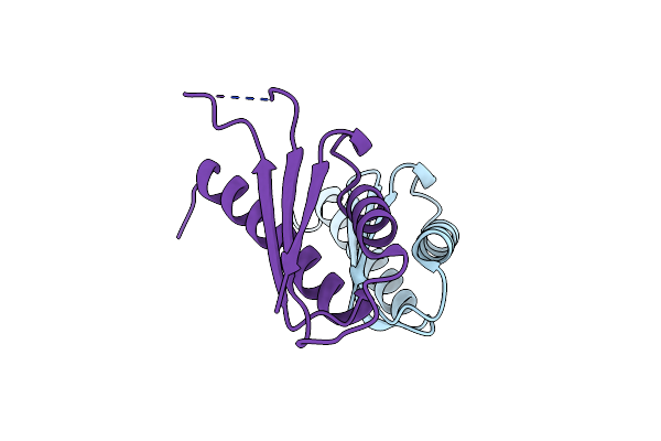
Crystal Structure Of The Type Iii Secretion System Pilotin-Secretin Complex Invh-Invg
Organism: Salmonella typhimurium (strain lt2 / sgsc1412 / atcc 700720)
Method: X-RAY DIFFRACTION Resolution:1.85 Å Release Date: 2020-09-16 Classification: PROTEIN TRANSPORT Ligands: SO4 |
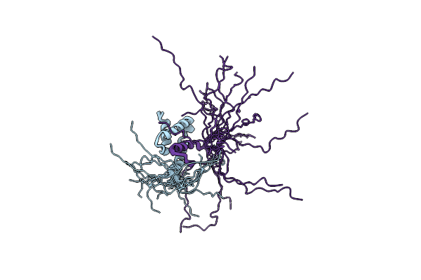 |
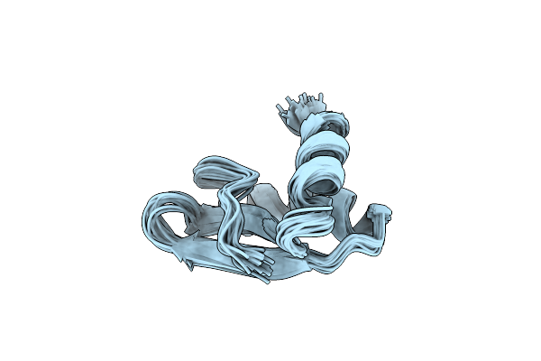
Structural Characterization Of The Type Iii Secretion System Pilotin-Secretin Complex Invh-Invg By Nmr Spectroscopy
Organism: Salmonella typhimurium (strain lt2 / sgsc1412 / atcc 700720)
Method: SOLUTION NMR Release Date: 2020-09-16 Classification: PROTEIN TRANSPORT |
 |
Organism: Mus musculus, Synthetic construct
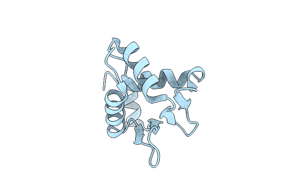
Method: X-RAY DIFFRACTION Resolution:2.00 Å Release Date: 2019-01-16 Classification: DNA BINDING PROTEIN Ligands: SO4 |
 |
Organism: Mus musculus, Synthetic construct
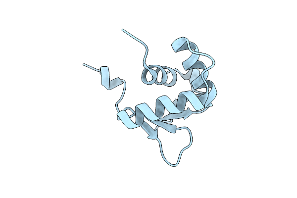
Method: X-RAY DIFFRACTION Resolution:2.35 Å Release Date: 2019-01-16 Classification: DNA BINDING PROTEIN Ligands: SO4 |
 |
Nmr-Based Structure Of The Fha-2 Domain From Mycobacterium Tuberculosis Abc Transporter Rv1747
Organism: Mycobacterium tuberculosis (strain atcc 25618 / h37rv)
Method: SOLUTION NMR Release Date: 2018-06-20 Classification: PROTEIN BINDING |
 |
Organism: Mycobacterium tuberculosis (strain atcc 25618 / h37rv)
Method: X-RAY DIFFRACTION Resolution:1.80 Å Release Date: 2018-06-20 Classification: PROTEIN BINDING Ligands: SO4 |
 |
Organism: Homo sapiens
Method: X-RAY DIFFRACTION Resolution:1.40 Å Release Date: 2017-02-22 Classification: DNA BINDING PROTEIN |
 |
Organism: Homo sapiens
Method: X-RAY DIFFRACTION Resolution:1.10 Å Release Date: 2017-02-22 Classification: DNA BINDING PROTEIN |
 |
Organism: Homo sapiens
Method: X-RAY DIFFRACTION Resolution:1.80 Å Release Date: 2017-02-22 Classification: DNA BINDING PROTEIN |
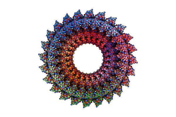 |
Organism: Salmonella enterica subsp. enterica serovar typhimurium str. lt2
Method: ELECTRON MICROSCOPY Release Date: 2015-01-14 Classification: STRUCTURAL PROTEIN |
 |
Organism: Salmonella typhimurium
Method: X-RAY DIFFRACTION Resolution:2.60 Å Release Date: 2014-12-03 Classification: PROTEIN TRANSPORT |
 |
Organism: Salmonella enterica
Method: SOLUTION NMR Release Date: 2014-10-29 Classification: CELL INVASION |
 |
Organism: Salmonella typhimurium
Method: X-RAY DIFFRACTION Resolution:3.20 Å Release Date: 2014-10-29 Classification: PROTEIN TRANSPORT |