
Search Count: 8
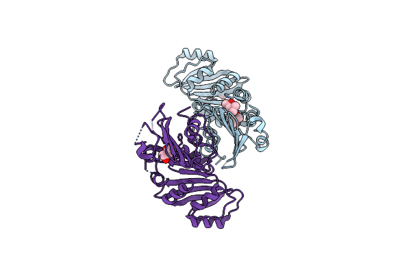 |
Organism: Treponema pallidum
Method: X-RAY DIFFRACTION Resolution:1.95 Å Release Date: 2010-10-27 Classification: MEMBRANE PROTEIN Ligands: MPD |
 |
Organism: Treponema pallidum
Method: X-RAY DIFFRACTION Resolution:2.39 Å Release Date: 2010-10-27 Classification: MEMBRANE PROTEIN Ligands: MPD |
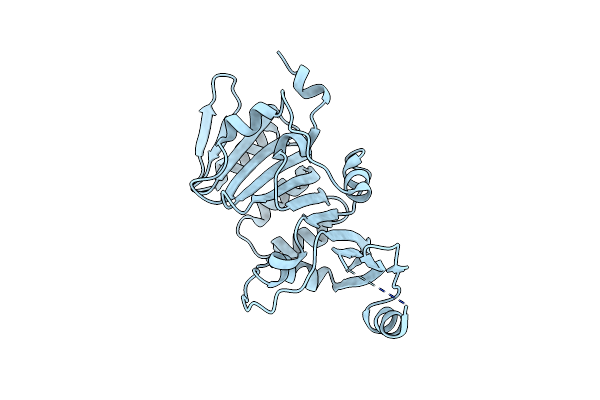 |
Organism: Treponema pallidum
Method: X-RAY DIFFRACTION Resolution:2.20 Å Release Date: 2010-10-27 Classification: MEMBRANE PROTEIN |
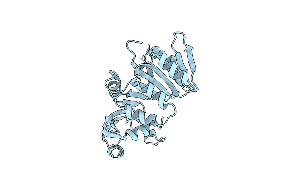 |
Organism: Treponema pallidum
Method: X-RAY DIFFRACTION Resolution:2.20 Å Release Date: 2010-10-27 Classification: MEMBRANE PROTEIN |
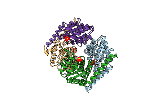 |
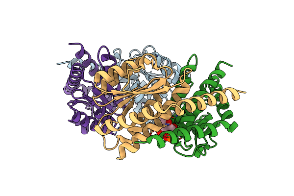
Organism: Pseudomonas aeruginosa pao1
Method: X-RAY DIFFRACTION Resolution:2.40 Å Release Date: 2007-12-18 Classification: ISOMERASE Ligands: SO4, CL |
 |
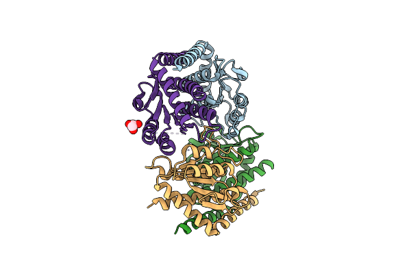
Crystal Structure Of Escherichia Coli Phosphoheptose Isomerase In Complex With Reaction Substrate Sedoheptulose 7-Phosphate
Organism: Escherichia coli
Method: X-RAY DIFFRACTION Resolution:2.80 Å Release Date: 2007-08-21 Classification: ISOMERASE Ligands: I22 |
 |
Organism: Escherichia coli
Method: X-RAY DIFFRACTION Resolution:1.95 Å Release Date: 2007-08-21 Classification: ISOMERASE Ligands: GOL |
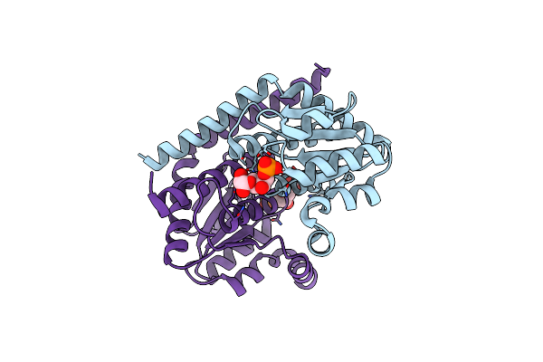 |
Crystal Structure Of Pseudomonas Aeruginosa Phosphoheptose Isomerase In Complex With Reaction Product D-Glycero-D-Mannopyranose-7-Phosphate
Organism: Pseudomonas aeruginosa
Method: X-RAY DIFFRACTION Resolution:2.30 Å Release Date: 2004-10-26 Classification: ISOMERASE Ligands: M7P |